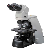
Chapter 2 Microscopy Operations
45
Epi-fluorescence
Microscopy
Preparation Focusing Microscopy
Epi-fluorescence
Microscopy Procedure
1234567891011121314 ■ ■ ■ ■ ■
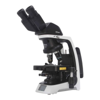
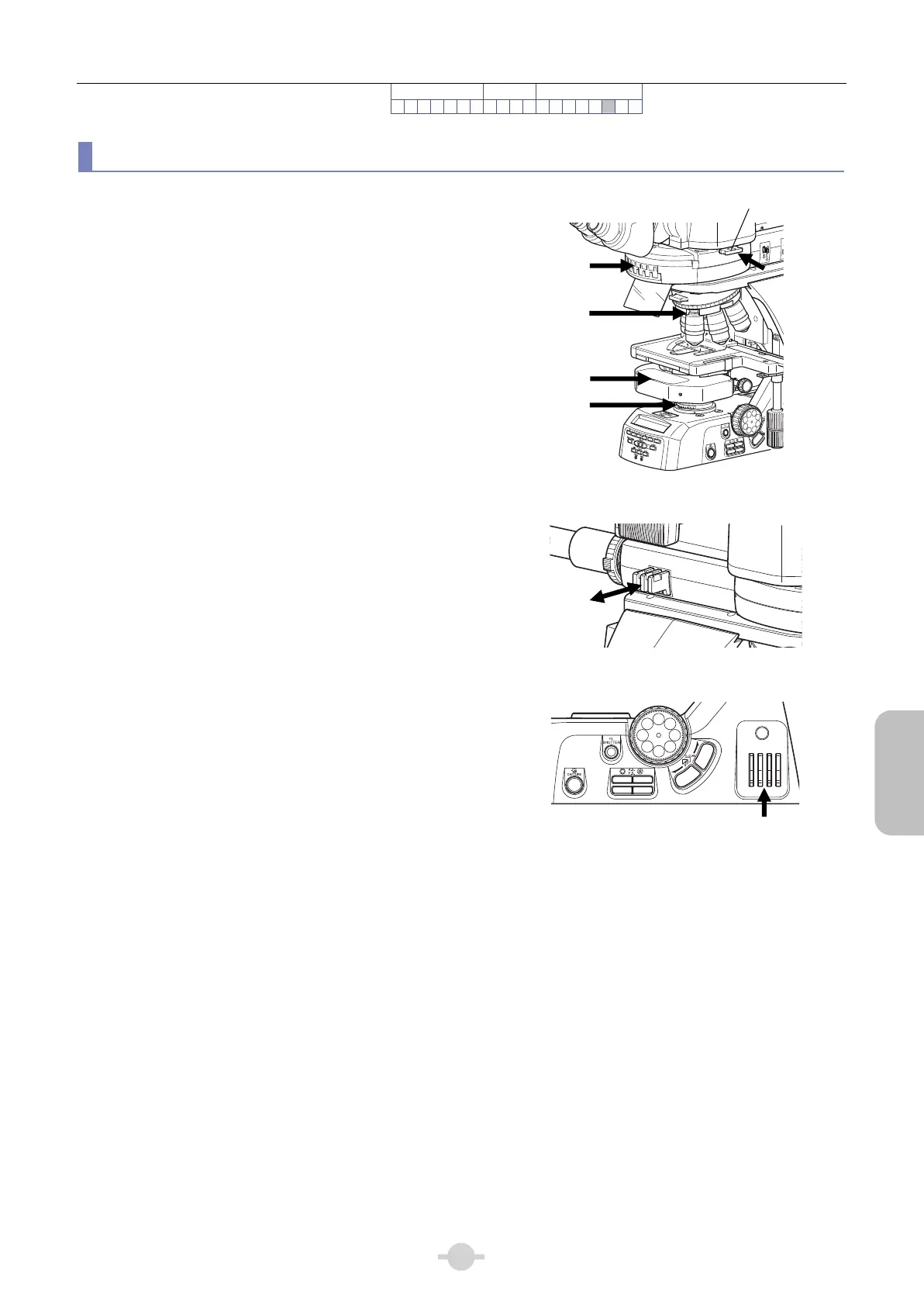
Concurrent Use of Epi-Fluorescence/Differential Interference Contrast Microscopy
By bringing the following optical elements into the optical
path, epi-fluorescence and differential interference
contrast can be simultaneously observed by microscope:
Filter cube for the epi-fluorescence cube turret
FL/DIC analyzer slider for the epi-fluorescence cube
turret
DIC slider on the objective side for the nosepiece
DIC module for the condenser
Polarizer for the polarizer unit
• When inserting the slider to the second click-stop
position in the DIC analyzer slider, the analyzer is
brought into the optical path. It is removed from the
optical path when the slider is pulled back to the
first click-stop position.
IN
OUT
1
2
A
λ
F. ST OP
EX.
2
3
4
5
6
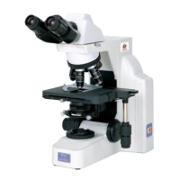
Concurrent use of epi-fluorescence/differential
interference contrast microscopy
ND4
ND8
ND16
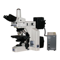
Brightness adjustment of fluorescent image
• If the diascopic image is not shown under
epi-fluorescence microscopy, press the dia-illumination
ON/OFF switch to turn on the lamp.
• Adjust the fluorescent image brightness with ND
filters in the epi-fluorescence attachment, and
differential interference contrast image brightness
using ND filters in the main body. Dia-illumination
should be sufficiently dimmed using the ND filter.
ND
8
ND
32
OUT
IN
NCB
11
Brightness adjustment of differential
interference contrast image
DIC slide
Filter cube
Polarize
DIC module
FL/DIC analyzer slide