10 Measuring Procedure
Instruction Manual Pentacam® / Pentacam HR® (G/70700/0109/en) 17 / 29
☞
Note
If the blue slit light is not visible, ensure that you have activated the [Slit Light] checkbox
on the "Scan" screen.
Darkening the room / dark sheet
Î If the lighting in the examination room has not been turned down or switched off,
use the dark sheet supplied to cover the patient and the Pentacam.
Adjustment
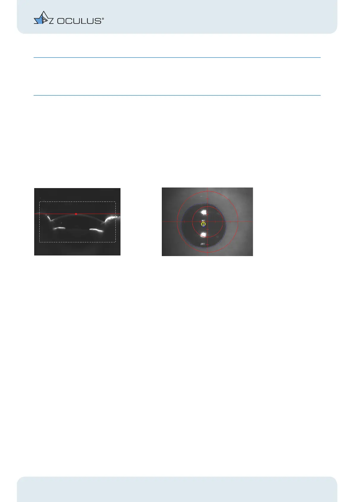
Î Move the cross slide towards the patient until the Scheimpflug image shows the
cornea of the eye that you are examining.
Fig. 10-4: Scheimpflug image (left) and pupil image (right)
The image is sharpest when the red dot coincides with the red line in the Sche-
impflug image.
Î Focus the pupil image by moving the joystick towards the Pentacam or away from it.
Î Adjust the left/right position of the Pentacam and its height setting.
Move the joystick to the left or right and rotate the joystick clockwise or anticlock-
wise.
The tentative final position of the camera is reached when the yellow dot is in the
centre of the crosshairs.
Î Ask the patient to widen his or her eye.
Fine adjustment
Î Make any fine adjustments required based on the information in the adjustment
window. To do so, move the joystick in the specified directions.
Example: Move the joystick to the right and forwards (towards the patient) and also
anticlockwise.