About the scanner Main characteristics
10
4522 207 12671/ * 2021-06-17
Pathology Scanner SG20 / SG60 / SG300
About the scanner
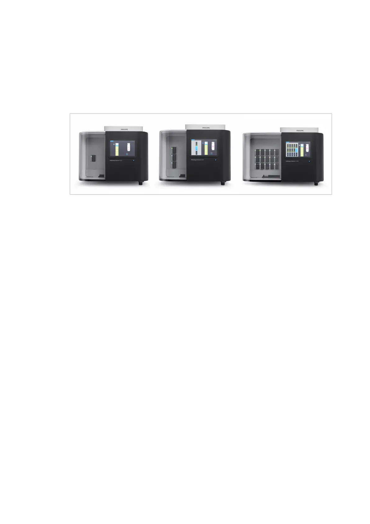
The Pathology Scanner SG20 / SG60 / SG300 is a dedicated scanner designed for scanning and
digitization of tissue glass slides. The scanner is connected to the Image Management System
(IMS).
Fig. 2: Pathology Scanner SG20 / SG60 / SG300
There are 3 different models with different slide capacities available:
• The SG20 can contain 1 rack with a maximum of 20 slides.
• The SG60 can contain 3 racks with a maximum of 20 slides each. When fully loaded, the
store contains 60 slides.
• The SG300 can contain 15 racks with a maximum of 20 slides each. When fully loaded, the
store contains 300 slides.
Main characteristics
The scanner has the following main characteristics:
• Fast scanning: Within 43 seconds
1
) a slide with a benchmark scan area of 15x15 mm
2
is
scanned with a resolution equivalent to a 40x magnification objective with a numerical
aperture of 0.75 (including handling and pre-scan, the total scan speed ≤ 62 seconds).
1
) For the Pathology Scanner SG20 the calculated scan time for a benchmark scan area as
above is about 101 seconds (including handling and pre-scan time, the total scan speed is
about 120 seconds).
• Tissue shape detection: The scanner adapts the scan region to the shape of the tissue. This
results in shorter scan times and decreased storage space (smaller image data files).
• Calibration check per slide: For each scan a check on the calibration is done. Only when the
check fails, the scanner will automatically start a calibration. This ensures that every slide is
scanned by a calibrated system and calibration is only triggered when truly necessary.
• Easy to use: The user only has to load and unload slides in the store, all other steps are fully
automated. The scanner automatically scans the slides, reads barcode information,
determines tissue location on the slides, auto-focuses on the tissue, scans the slides at high
resolution and transfer the images to the IMS.
• Continuous loading/unloading: The user can load and unload slides without interrupting
the scanning process. After loading new slides, the scanner will schedule the scanning of the
new slides while continuing with its current job.
2