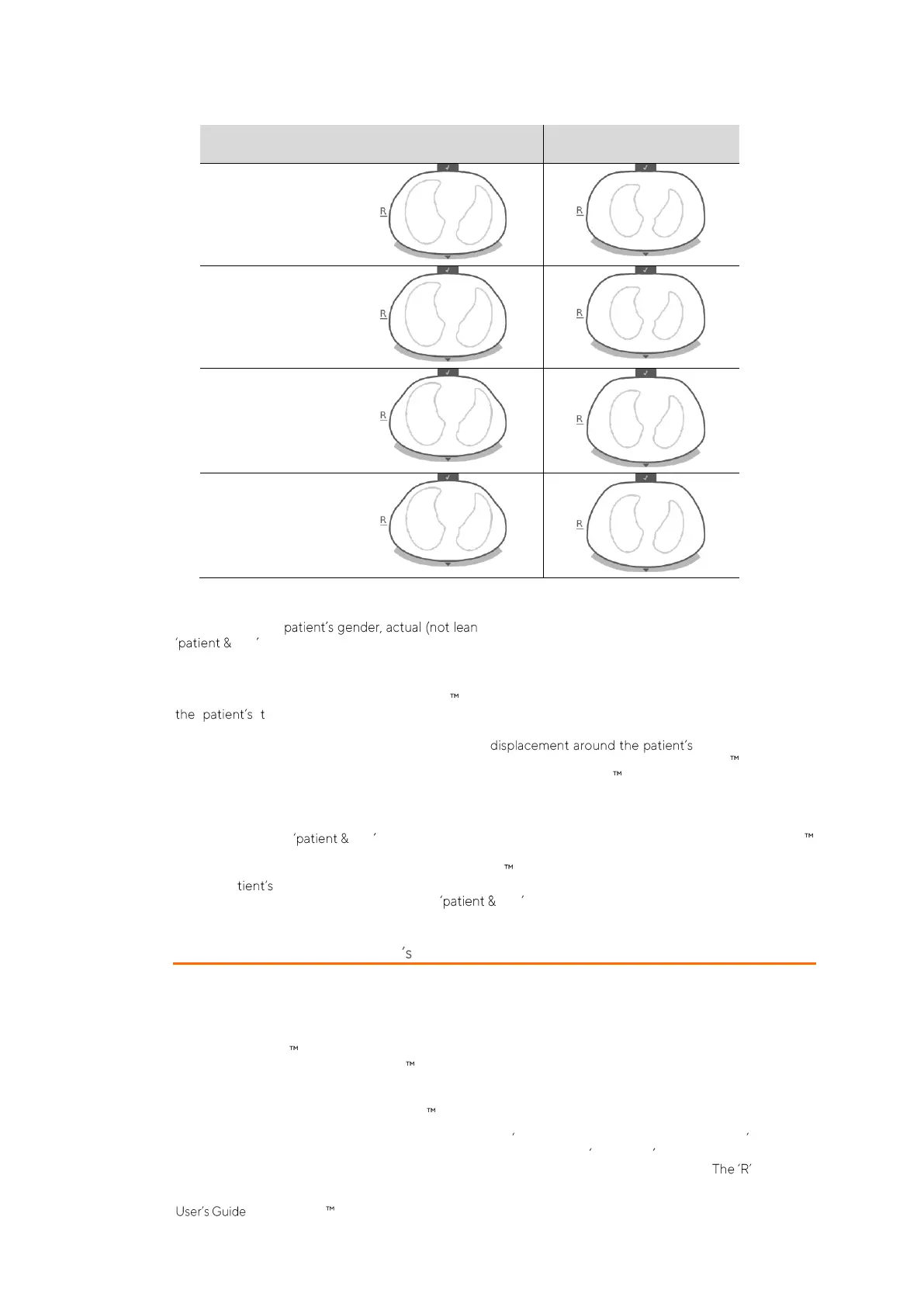
To select the thorax and lung model being best adapted to an individual patient in Adults/Children
configuration, the or ideal) body weight, and height must be entered in the
belt area of ScoutView (Figure 6-7) when initiating monitoring for a new patient. In Neonates/Infants
configuration, only one CT-derived thorax and lung model is used and, consequently, the
Thorax and Lung
Contours
used for the various
EIT images
are identical for all patients.
The thorax and lung models used by the LuMon System also assume specific positions of the 32 electrodes on
horax. In order to obtain optimal data, it is therefore very important to apply belts of
recommended size to the patient properly (8) and to check belt positioning regularly (at least every 8 hours)
during monitoring (8.4). To avoid an inadvertent belt thorax, which would
result in undefined electrode positions, belts for adults and children (SensorBelts/LuMon Belts Adult
(5.3.1/5.3.2), have shoulder strips. Belts for neonates and infants (LuMon Belts Neo (5.3.3)), do not have
shoulder strips and it is therefore possible that in neonates and infants the belt is inadvertently rotated or
displaced around the thorax from its optimal position. In order to assess the electrode positions on the thorax of
neonates and infants as accurately as possible, it is therefore important to measure and enter the so-called belt
displacement in the belt area of ScoutView (Figure 6-7) when applying a SensorBelt (8.1) or LuMon
Belt (8.2, 8.3), check it regularly (at least every 8 hours) and eventually update it during monitoring (8.4).
Moreover, in Neonates/Infants configuration, the LuMon System also factors in the size of the connected belt
and the pa full underbust girth to determine the electrode positions. The full underbust girth is also to be
measured and entered by the operator in the belt area of ScoutView (Figure 6-7) when applying the
belt and initiating monitoring in Neonates/Infants configuration (8.3).
9.2 Accounting for the patient position
It is well known that physiological and pathophysiological phenomena are influenced by gravity (e.g. [6]). In
order to facilitate the user with the possibility to better account for the influence of gravity on lung mechanics
and ventilation distribution when evaluating monitored data displayed in real-time or retrospectively in the
trends, the LuMon System continuously measures, visualizes and records patient position (
rotation
and
inclination
). Furthermore, the LuMon System considers the measured
rotation
when calculating the
Dependent Silent Spaces
(DSS), the
Non-Dependent Silent Spaces
(NSS) as well as the vertical and horizontal
components of the
Center of Ventilation
(CoV(v) and CoV(h)). To this aim a position sensor is embedded in
SensorBeltConnectors (5.2.1) and in LuMon Belts (5.3.2/5.3.3).
To indicate the measured
rotation
, all
EIT images
and the Belt-connection/Skin-contact/Rotation icon in the
Status Bar (6.6.1) rotate with the patient.
Inclination
is indicated with the Inclination icon in the status bar (6.6.1)
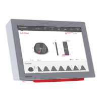
and on ScoutView (6.2). Figure 9-2 illustrates how patient position is indicated on ScoutView. at the side


 To Next Page