It is important to note that, thanks to the position sensor, DSS and NSS as well as the coordinates CoV(v) and
CoV(h) are always automatically defined if
rotation
changes, thereby allowing to directly assess the influence of
ventilation distribution. Only in supine position, the coordinates CoV(v) and
CoV(h) used by the LuMon System are ali body
directions typically used in literature [3], [8] and illustrated in black in Figure 9-17, namely the right-left and
ventrodorsal coordinates, CoV(rl) and CoV(vd) respectively.
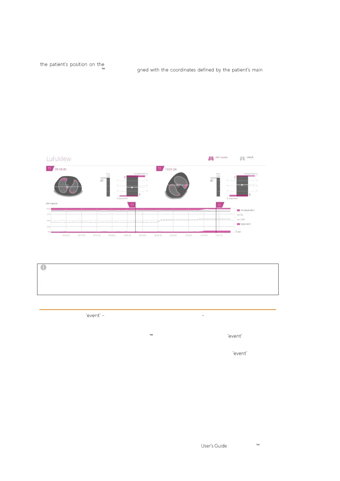
The FLS bar (in the center of Figure 9-16) represents the percentage of FLS with respect to the total lung area,
and the
Silent Spaces
bar (on the right of Figure 9-16) represents the respective percentages of NSS and DSS
(FLS, DSS and NSS together totaling 100%). Here CoV is also displayed as a whitish dot for contextualization,
with its vertical coordinate represented on the right vertical scale with 0% on the top (its horizontal coordinate
being also displayed but without a scale).
These percentages and the vertical coordinate of CoV are also shown in the corresponding trend graph as
illustrated in Figure 9-18. Two time points, T1 and T2, are set for comparison. DSS, NSS, CoV(v) and FLS are
shown in the trend graph in percentage values. The values of NSS and CoV(v) can be visualized considering 0%
on the top, those of DSS and FLS considering 0% on the bottom.
Figure 9-18: Example of a Silent Spaces trend graph.
Finally, yet importantly, be aware that the subdivision of
Silent Spaces
into
Dependent Silent Spaces
(DSS) and
Non-Dependent Silent Spaces
(NSS) as well as the determination of the vertical and horizontal components of
the
Center of Ventilation
(CoV(v) and CoV(h)) depend on displayed
rotation
(see also Figure 9-17).
CAUTION
To avoid misinterpretation of results be aware that the
Dependent Silent Spaces
, the
Non-Dependent Silent
Spaces
as well as the vertical and horizontal components of the
Center of Ventilation
(CoV(v) and CoV(h)) are
less meaningful, if the gravity vector is more or less perpendicular to the examined thorax cross-section being
defined by the
belt plane
. This is, for example, the case, if the patient is standing or sitting upright.
9.11 Operator events and screenshots
Pressing the button which is available on all views (Figure 6-5) will mark an operator event. As
illustrated in Figure 9-19 dotted black vertical lines with triangles at both ends identify operator events in the
trends and on the
Plethysmogram
(9.4).
If a USB memory device is connected to the LuMon Monitor when pressing the button a screenshot of
the Graphical User Interface (at the time the button is pressed) is additionally stored on the USB memory
device. Screenshots are stored in standard JPG image format and can be read by standard image viewer
software. The filename of a screenshot corresponds to the date/time when the button was pressed
(yyyymmddhhmmss.jpg).


 To Next Page