Do you have a question about the Sirona Galaxis and is the answer not in the manual?
Explains symbols and character formats used in the manual.
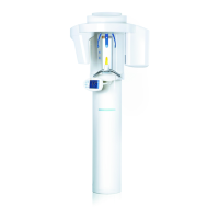
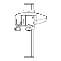
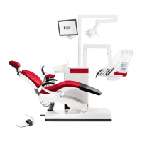
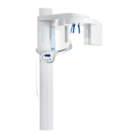
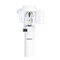
Covers intended use, system components, and important notices for GALAXIS.
Details the reduced functionality when using GALAXIS with model version GAX5.
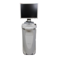
Lists the minimum hardware and software requirements for the RCU.
Outlines the minimum hardware and software specifications for the PC running GALAXIS.
Specifies the requirements for network connectivity within the GALAXIS system.
Describes the layout of workspaces, including title bar, menu, toolbar, views, and tabs.
Explains the classical panoramic view and its associated examination window.
Details the workspace displaying axial, coronal, and lateral planes along with the 3D view.
Describes the workspace for viewing the posteroanterior or anteroposterior cephalometric projection.
Details the workspace for viewing the lateral cephalometric projection.
Explains the workspace for an enlarged view of a specific area within the examination window.
Covers the reconstruction of the panoramic view and its internal examination window.
Explains how the volume is displayed as a 3D object and its interaction features.
Describes the straight slice oriented longitudinally to the examination window.
Explains the slices oriented perpendicular to the mandibular arch.
Details the horizontally positioned slices displayed at the same height as horizontal lines in other views.
Describes the frontal slices of the volume, part of the Radiology workspace.
Explains the frontal slices of the volume, part of the Radiology workspace, and rotation options.
Allows transferring current views or examinations to SIDEXIS XG as 2D images.
Explains how to print the current view or the complete examination.
Describes methods for moving through slices using mouse buttons and dedicated buttons.
Details the default cursor mode and how to switch to it.
Explains the mouse adjuster for simultaneously adjusting image brightness and contrast.
Describes how to use the "Hand" mode to move the enlarged slice plane.
Explains how to position the crosshair at a specific location with a single click.
Details how to reset all views and settings to their default values.
Covers functions for measuring lengths and angles within views.
Guides on reconstructing a panoramic view if the default quality is insufficient.
Explains how to increase or decrease the size of any view using zoom functions.
Describes how to change gamma correction to modify the display of shades of gray.
Explains how to create, select, edit, delete, and manage findings as bookmarks.
Details the process for reconstructing posteroanterior or anteroposterior cephalometric views.
Guides on reconstructing a lateral cephalometric view from the volume.
Explains how to create a detailed reconstruction of a specific region.
Describes how to show or hide orientation lines and findings in the views.
Covers customize settings for closing GALAXIS and adjusting view options.
| Model | Galaxis |
|---|---|
| X-ray tube current | 7 mA |
| Focal spot size | 0.8 mm |
| Image receptor | Digital sensor or film |
| Manufacturer | Sirona |
| X-ray generator voltage | 60-70 kV |