61 23 488 D 3474
22 D 3474.208.01.06.02 04.2010
Views Sirona Dental Systems GmbH
Panoramic view Operator's Manual GALAXIS
7 Views
7.1
Panoramic view
Explanation The panoramic view is reconstructed based on a U-shaped region correspon-
ding to the mandibular arch.
● The examination window [ ➙ 22] is located inside the panoramic view.
● The U-shaped region is displayed in the axial plane of the corresponding
workspace.
● The displayed panoramic view can be reconstructed by altering the U-
shaped region (see chapter: Reconstructing a panoramic view [ ➙ 34].)
7.1.1 Examination window
Explanation The examination window is located inside the panoramic view.
The examination window displays a thin, vertical slice of the volume along the
mandibular arch.
Configuration
Position The position of the examination window is shown by the curved line in the axi-
al slice of the corresponding workspace, as well as by the vertical line in the
transversal views and in the longitudinal view.
NOTICE
The subjective image impression of the reconstructed panoramic view is not
necessarily comparable to the conventional blurring technique (e.g. OR-
THOPHOS) due to the dental volume tomography technique used.
The detail accuracy has been verified by numerous scientific publications.
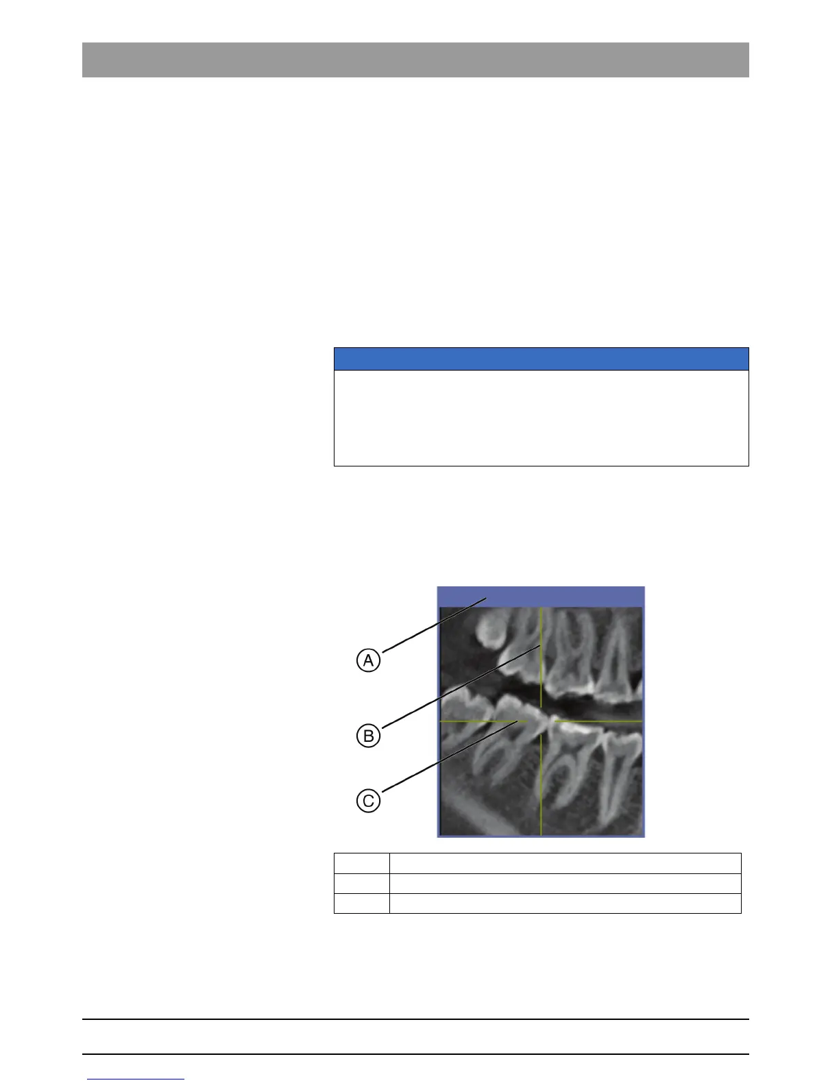
A Title bar.
B Position of the transversal view (TSA) [ ➙ 25].
C Position of the axial plane [ ➙ 27].