61 23 488 D 3474
12 D 3474.208.01.06.02 04.2010
Creating 3D X-ray images Sirona Dental Systems GmbH
Operator's Manual GALAXIS
5 Creating 3D X-ray images
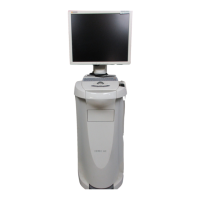
Explanation The 3D X-ray images are created via the SIDEXIS XG exposure readiness
function and managed in the SIDEXIS database parallel to other X-ray ima-
ges (IO "Intraoral", XP "Panoramic", XC "Ceph", etc.).
At the same time, the 3D X-ray images are stored on the
"3D"
tab card.
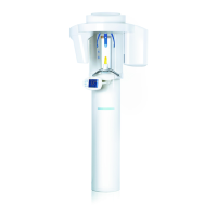
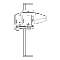
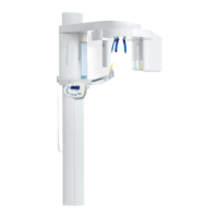
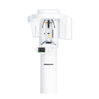
Required documentation The operation of the GALILEOS X-ray device is explained in the
"GALILEOS
Operating Instructions"
.
The operation of SIDEXIS XG software is explained in the
"SIDEXIS XG Ope
-
rator's Manual"
.
Creation 1. Start SIDEXIS XG (see
"SIDEXIS XG Operator's Manual"
).
2. Select a patient.
3. Press the 3D Scan button.
4. Press the release button.
After a few minutes, the icon for the 3D X-ray image is displayed on the SI-
DEXIS XG desktop.
The 3D X-ray image is now managed in the SIDEXIS database in the same
way as any other X-ray image.
CAUTION
X-rays
Make sure that all locally applicable radiation protection regulations are ob-
served! Also make sure that the application information provided in the
"GA
-
LILEOS Operating Instructions"
is observed (especially regarding patient
positioning and the start of exposure)!