18
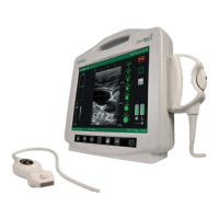
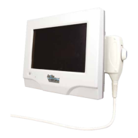
Instructions For Use
Step 6: Evaluate External ECG waveform
- Refer to the Bard Access Systems’ catheter Instructions for Use.
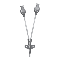
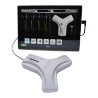
Step 7: Perform Initial Magnet Tracking Calibration
- Calibrate the Sherlock 3CG™ TCS by selecting CALIBRATE [ ] prior to setting up the sterile field to ensure there is no environmental interference.
Tip: If calibration fails, remove any items that may be causing magnetic interference (e.g. active motor driven equipment, monitor leads, cell phones, name
tags, jewelry, etc.).
Step 8: Prepare Catheter Sterile Field
- Refer to the Bard Access Systems’ catheter Instructions for Use.
Step 9: Access the Vein
- Refer to the Bard Access Systems’ Ultrasound System Instructions and Bard Access Systems’ catheter Instructions for Use.
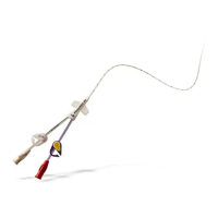
Step 10: Attach Catheter Stylet to Fin Assembly
- Refer to Bard Access Systems’ catheter Instructions for Use.
Step 11: Perform Final Magnet Tracking Calibration
- Ensure the catheter tip is at least 12 inches (30 cm) away from the sensor before calibrating.
- Select CALIBRATE [ ] immediately prior to catheter insertion.
- Once calibration is complete, ask the patient to remain still and do not reposition the patient.
- Refer to Bard Access Systems' catheter Instructions for Use for catheter insertion.
Step 12: Insert Catheter
- Refer to Bard Access Systems’ catheter Instructions for Use for catheter insertion.
Step 13: Catheter Tip Guidance and Positioning
- Refer to Bard Access Systems’ catheter Instructions for Use for catheter insertion.
- Initially a searching magnifying glass will indicate that the stylet tip is outside the sensor range.
- Use a slow steady motion while advancing the catheter.
Magnetic Navigation
- As the stylet tip approaches the sensor, an icon appears at the edge of the screen indicating the approach of the stylet tip.
- When the stylet is under the sensor, the stylet and depth icons will display the location, orientation, and depth of the stylet in relation to the sensor.
- Advance the catheter slowly to achieve optimal performance (1 cm per second). There may be a slight delay from the time the catheter is moved until the
stylet icon moves on the display. Advancing the catheter too quickly may result in erratic movements of the stylet icon on the display.
- Insert the catheter until the magnetic navigation shows the stylet moving consistently downward.
- Continue to slowly advance catheter until the catheter is inserted to the external measurement as determined in the Bard Access Systems’ catheter
Instructions for Use.
- Select [ ] to minimize the magnetic navigation window and freeze/save the current ECG waveforms in the reference screen.
Note: select the minimized magnetic navigation window to return to a maximized state
ECG Positioning
- In patients with a distinct P-wave, the P-wave will increase in amplitude as the catheter approaches the cavo-atrial junction. As the catheter advances into
the right atrium, the P-wave will decrease in amplitude and may become biphasic or inverted.
Note: If the intravascular ECG waveform is not displayed, flush the catheter with saline. If the problem continues, check the stylet-to-fin connection.
To freeze and compare ECG waveforms select [ ] to copy ECG waveforms in the reference window.
Note: Pause to let the rhythm settle before freezing the reference ECG waveforms.
- For final catheter positioning, refer to Bard Access Systems' catheter Instructions for Use.
Step 14: Complete Catheter Placement
- Complete catheter insertion, securement and remaining procedure according to the catheter Instructions for Use and facility protocol.
 Loading...