44 Software User Guide Rev. 1.0 Kolibri cranial/ENT Ver. 2.7
Tracking System
Camera View
Windows
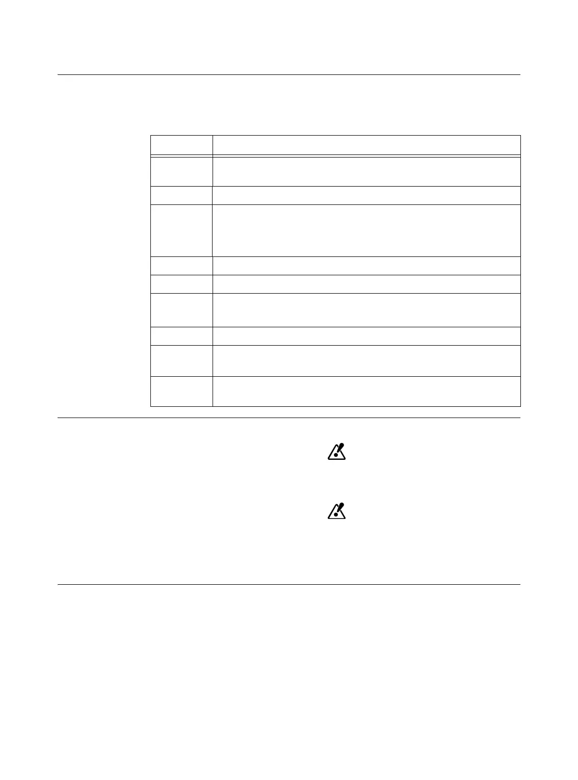
The camera view windows display dots which represent the marker sphere geometries of the
reference arrays and navigated instruments. The color-coding of the dots is specific to each
reference array and instrument. You can use the camera view windows to check whether an
instrument or array is visible to the camera
Instrument Display
Color blind users may not be able to clearly distinguish color differences of the
represented instruments.
Make sure that the instrument marker arrays are not displayed too closely to-
gether and that they do not block other markers from being detected by the
camera. Otherwise the system may not be able to differentiate between instru-
ments, resulting in incorrect instrument navigation.
Dot Color Represented Element
Red
Reference array (e.g.,
Mayfield Reference Array
) if the patient has been
registered
Yellow Calibrated
StarLink Instrument Adapter
marker spheres or the
ICM
Orange
Calibration devices
(during instrument calibration):
•
ICM
•
Reference Array
Green Pointer or
Softouch
Blue Microscope adapters
Pink
•
Disposable Biopsy Needle Type A
and
Biopsy Alignment Array
•
VarioGuide
White Uncalibrated instruments or reference array before patient registration
Gray
(filled)
Marker spheres of an array are visible to both camera lenses, but cannot
be assigned to a specific instrument
Gray
(open)
Array is only visible to one camera lens and cannot be interpreted
 Loading...