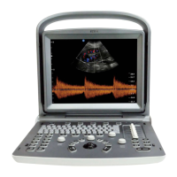
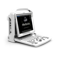
ECO 3 EXPERT Ultrasound Diagnostic System
Page 43 / 191
5.5 Display Mode
Display Mode: B, 2B, 4B, M, B/M, they can be shifted by the mode key.
5.5.1 B Mode
Press [B] Mode key, and display the single B Mode image, B Mode is the basic mode for
two-dimensional scanning and diagnosis.
5.5.2 2B Mode
Press [2B] to display double B mode images side by side. One image is in real-time status; the
other is in frozen status. The real-time image has start scan marker and ruler marker .Press 2B
button in 2B mode, the original active image is frozen while the original frozen image is activated.
In frozen status, Press [2B] to choose a B mode image to be activated when unfreezing the image.
5.5.3 4B Mode
Press [4B] button to enter into 4B mode, the screen will display four B mode images side by side,
but only one image is in real-time status. Pressing it again can switch the real-time status among
four images.
In frozen status, Press [4B] to choose a B mode image to be activated when unfreeze the image.
5.5.4 B/M Mode
Press [M] button, a real time B-mode image and a real-time M-mode image will be displayed at the
same time. And a sample line will appear in the B-mode image area, which indicates the active
sample position for M image on the B image area. Move the sampling line by trackball.
5.5.5 M Mode
Press [M] key again, B mode image will disappear; M mode image is still active on the whole
screen. M mode image stands for the tissue movement status at the sampling line. The M mode
image varies with time, so it is mainly used for cardiac applications.
5.5.6 PW Mode
Doppler is intended to provide measurement data concerning the velocity of moving tissues and
fluids. PW Doppler lets you examine blood flow data selectively from a small region called the
 Loading...