Do you have a question about the Chison ECO 3 EXPERT and is the answer not in the manual?
Provides an overview of the ultrasound system and its intended use.
Details on how to contact CHISON for support and information.
General measures to ensure the safety of both operator and patient.
Details on protection against electric shock and grounding requirements.
Explanation of symbols and labels found on the device panel.
Specifications for devices used within the patient environment and their compatibility.
Guidelines for safe use of diagnostic ultrasound regarding thermal and mechanical effects.
Usage guidelines for safe operation and understanding scanning parameters.
Precautions for the safe use and storage of Li-ion batteries.
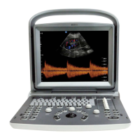
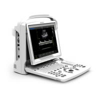
Visual description of the ultrasound system's console layout.
Dimensions and weight of the main ultrasound unit.
Illustrations showing the system from front, side, and rear views.
Overview of the system's various imaging modes and capabilities.
Steps and requirements for installing the ultrasound system.
Description of the alphanumeric keys used for inputting patient data.
Explanation of various function keys and knobs for system operation.
Details on the central control panel elements like trackball and navigation keys.
Description of keys used to select different image display modes.
Functions for adjusting image quality and display parameters.
Steps for inspecting and powering on the ultrasound system.
Procedure for selecting the probe and examination type.
How to input and manage patient information for examinations.
Description of the elements displayed on the ultrasound image interface.
Explanation of different screen display modes like B, 2B, 4B, B/M, M, PW.
Controls and settings for optimizing B mode image quality.
Detailed adjustments available within the B mode image menu.
Settings for optimizing Pulsed Wave (PW) Doppler image parameters.
How to activate and exit the full screen image display.
Procedures for adding and managing text comments on images.
How to select and place body marks to indicate probe position.
Instructions for adding and adjusting directional arrows on the image.
Principles and procedures for storing and recalling images and cine loops.
Interface for browsing and managing stored images.
Functionality for searching and managing patient information archives.
Creating, saving, and printing examination reports.
Setup and management of DICOM services like storage and worklist.
Details on using the trackball and ENTER key for measurements.
Explanation of Distance, Ellipse, and Trace measurement techniques in B mode.
Quick measurement options available in B mode.
Standard measurement procedures for B mode in various categories.
Specific measurement protocols for abdominal examinations.
Measurement parameters and formulas for obstetric examinations.
Measurement procedures specific to pediatric examinations.
Measurement protocols for gynecological examinations.
Procedures for measuring small anatomical parts.
Vessel measurement techniques in B mode.
Measurement protocols for urological examinations.
Measurement procedures specific to cardiac examinations.
Standard measurements for M and B/M modes.
General measurement functions applicable to M mode.
Abdomen-specific measurements in M mode.
Obstetrics-specific measurements in M mode.
Gynecology-specific measurements in M mode.
Cardiac measurements performed using M mode.
Urology-specific measurements in M mode.
Small parts measurements in M mode.
Pediatric-specific measurements in M mode.
Methods for performing measurements in Pulsed Wave (PW) mode.
Quick measurement options for PW Doppler.
General measurement procedures for PW Doppler mode.
Abdomen-specific measurements using PW Doppler.
Obstetrics-specific measurements using PW Doppler.
Gynecology-specific measurements using PW Doppler.
Cardiac measurements using PW Doppler.
Vascular measurements using PW Doppler.
Urology-specific measurements using PW Doppler.
Small parts measurements using PW Doppler.
Pediatric-specific measurements using PW Doppler.
Configuration of general system parameters like hospital name and language.
Customization of measurement units, formulas, and display options.
Configuration of annotation library, comments, and arrows.
Customization of body marks for indicating probe scanning position.
Defining and customizing exam modes, including comments and body marks.
Setting up report layout and diagnostic templates.
Setup and management of DICOM services like storage and worklist.
Configuration of network connections, IP setup, and storage.
Displaying system information and procedures for software/hardware updates.
Guidelines for safely cleaning the ultrasound system.
Procedures for regular safety checks and addressing malfunctions.
Table listing common malfunctions, their reasons, and measures.
Overview of the ultrasound probes and their specifications.
Instructions for inspecting, cleaning, disinfecting, and storing probes.
Guidelines for connecting, using, and handling ultrasound probes.
Information on manufacturer's service responsibilities and product life.
| Brand | Chison |
|---|---|
| Model | ECO 3 EXPERT |
| Category | Diagnostic Equipment |
| Language | English |