Chapter 2 Microscopy
22
Return to the bright-field microscopy under the episcopic illumination.
1 Pull the polarizer/analyzer unit to the first
stop-click position and remove the
polarizer/analyzer from the optical path. (See
3.13.1.)
2 Both the λplate and the polarizer/analyzer unit are
removed from the optical path. (See 3.14.1.)
3 Detach the DIC slider to remove the DIC prism
from the optical path. (See 3.15.1 or 3.16.2.)
4 Turn the brightness control dial to adjust the
brightness of the episcopic illumination. (See
3.2.1.)
5 Adjust the brightness with the desired ND filter in
the filter turret. (See 3.10.)
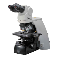
2.5 Epi-fl Microscopy
Attach the items required for the epi-fl microscopy to the microscope. (See Table 2.1.)
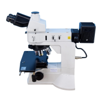
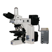
To perform the epi-fl microscopy using the C-HGFI/C-HGFIE HG Precentered Fiber Illuminator as the
external light source, screw the compensation filter supplied with the fiber adapter into the MA2-FL
fluorescent unit. (Refer to 4.15.1.)
Find the target and focus on the sample by BF/DF microscopy under the episcopic
illumination. (See Pages 16 to 17, and 19.)
Set the microscope for the epi-fl microscopy.
1 Turn the revolving nosepiece to place the
objective of a desired magnification into the optical
path. (See 3.9.2.)
2 Turn the BD field changeover lever to the “BF
(bright-field)” position. (See 3.3.)
3 Push the attached fluorescent unit into the second
click-stop position and place the fl filter into the
optical path (See 3.19.1.)
4 Turn the brightness control dial to adjust the
brightness. (See 3.2.1.)
5 Adjust the brightness with the desired ND filter in
the filter turret. (See 3.10.)
3-4
3-5
3-3
3-2
3-1