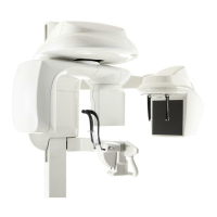
Acquiring a Carpus Image
CS 9300C User Guide (SM750)_Ed 01 5–27
Acquiring a Carpus Image
Through radiological analysis of ossification to the carpal bone, you can assess the
growth or maturity rate of a pediatric patient.
Before acquiring a carpus image, check that you have:
Reset the unit rotative arm to the start position for patient to enter the unit.
Selected the patient record.
Accessed the Imaging window.
Accessed the Cephalometric Acquisition interface.
Preparing the Unit and Setting the Acquisition Parameters
for the Pediatric Patient
To acquire a carpus image, follow these steps:
1. Position the head clamps manually for a frontal AP exam.
2. In the Cephalometric Acquisition interface, click the Program button to access the
Program pane. In the Program pane:
The for a frontal AP exam is active.
Click for a carpus exam.
Select the 30 x 30 acquisition format.
3. Click the Patient button to access the Patient pane. Select the pediatric patient type.
4. If the default parameter setting is not adapted to your pediatric patient type, click the
Parameter button and select the appropriate parameters. To save the new parameter
settings as the default settings, click
and select Memorize settings.
IMPORTANT
You must position the head clamps manually because they
are not positioned automatically from the Program pane exam
type selection. In this case, the relevant exam type selection
icon becomes active.