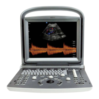
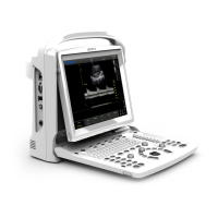
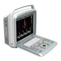
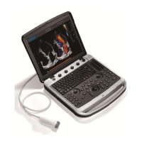
ECO 6 Digital Color Doppler Ultrasound System
12
Where a is the attenuation coefficient in dB cm-1 MHz-1, f is the transducer center frequency, and z is the
distance along the beam axis between the source and the point of interest.
De-rating factor RF for the various distances and frequencies with attenuation coefficient 0.3dB cm-1 MHz-1
in homogeneous soft tissue is listed in the following table. An example is if the user uses 7.5MHz frequency,
the power will be attenuated by .0750 at 5cm, or 0.3x7.5x5=-11.25dB. The De- rated Intensity is also referred
to as ‘.3’ at the end (e.g. Ispta.3).
I’=I*RF Where I’ is the intensity in soft tissue, I is the time-averaged intensity measured in water.
Tissue Model:
Tissue temperature elevation depends on power, tissue type, beam width, and scanning mode. Six models are
developed to mimic possible clinical situations.
Soft tissue:
Describes low fat content tissue that does not contain calcifications or large gas-filled spaces.
Scanned: (auto-scan)
Refers to the steering of successive burst through the field of view, e.g. B and color mode.
Unscanned:
Emission of ultrasonic pulses occurs along a single line of sight and is unchanged until the transducer is moved
to a new position. For instance, the PW and M mode.
WWW.CFS.IT - CFS PRODOTTI MEDICALI SRL
 Loading...