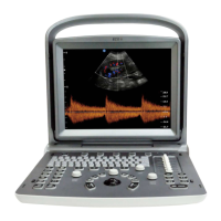
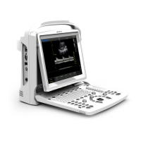
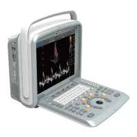
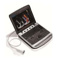
ECO 6 Digital Color Doppler Ultrasound System
36
5.5.14 Biospy and Super Needle
1. How to enter into Biopsy
Activate the [Biopsy] and press the [MENU] knob to show or hide biopsy line.
2. How to adjust the biopsy
After the biopsy line shows, press the [ENTER] key to activate the adjustment function of biopsy line,
horizontal rolling the trackball can translate the biopsy line, vertical rolling the trackball can adjust the line
angle, press the [UPDATE] key to set the default biopsy line position.
3. Super Needle
Super needle is used for enhancing the needle image in the B mode image. After turning on the super needle,
super needle and needle angle function will be active and user can adjust the needle angle to optimize the
image for needle only (The angle is 5 degree per step.).
5.5.15 PW Mode
PW Doppler is intended to provide measurement data concerning the velocity of moving tissues and fluids.
PW Doppler lets you examine blood flow data selectively from a small region called the Sample Volume.
The X axis represents time while the Y axis represents velocity in either a forward or reverse direction.
PW Doppler is typically used for displaying the speed, direction, and spectral content of blood flow at selected
anatomical sites.
PW Doppler can be combined with B mode for quick selection of the anatomical site for PW Doppler
examination. The site where PW Doppler data is derived appears graphically on the B mode image (Sample
Volume Gate). The Sample Volume Gate can be moved anywhere within B mode image.
PW mode Exam Procedure:
Get a good B mode image. Press [C] key to help to locate the vessel you wish to examine.
Press [D] key to display the sample volume cursor and gate.
Position the sample volume cursor by moving the Trackball left and right. Position or re-size the sample
volume gate by moving the Trackball up and down, then press [ENTER] key.
Press [UPDATE] key to display PW Doppler spectrum and the system will run in combined B+Doppler
mode. The Doppler signal can be heard through the speakers.
Optimize the PW Doppler spectrum as necessary.
Ensure that the sample line is parallel to the blood flow.
Press [FREEZE] key to hold the trace in cine memory and stop imaging.
Perform measurements and calculations, as necessary.
Record results with your recording devices.
Press [FREEZE] key to resume imaging.
Repeat the above procedure until all relevant flow sites have been examined.
Replace the probe in its respective holder.
When entering Duplex mode for the first time, the Doppler spectrum is not activated. The Doppler Sample
Volume appears in the default position, and the B mode image or 2D (either B or Color) mode are active.
Moving the Trackball will change the Sample Volume position. Press the [ENTER] key to toggle the Trackball
WWW.CFS.IT - CFS PRODOTTI MEDICALI SRL