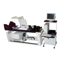
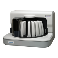
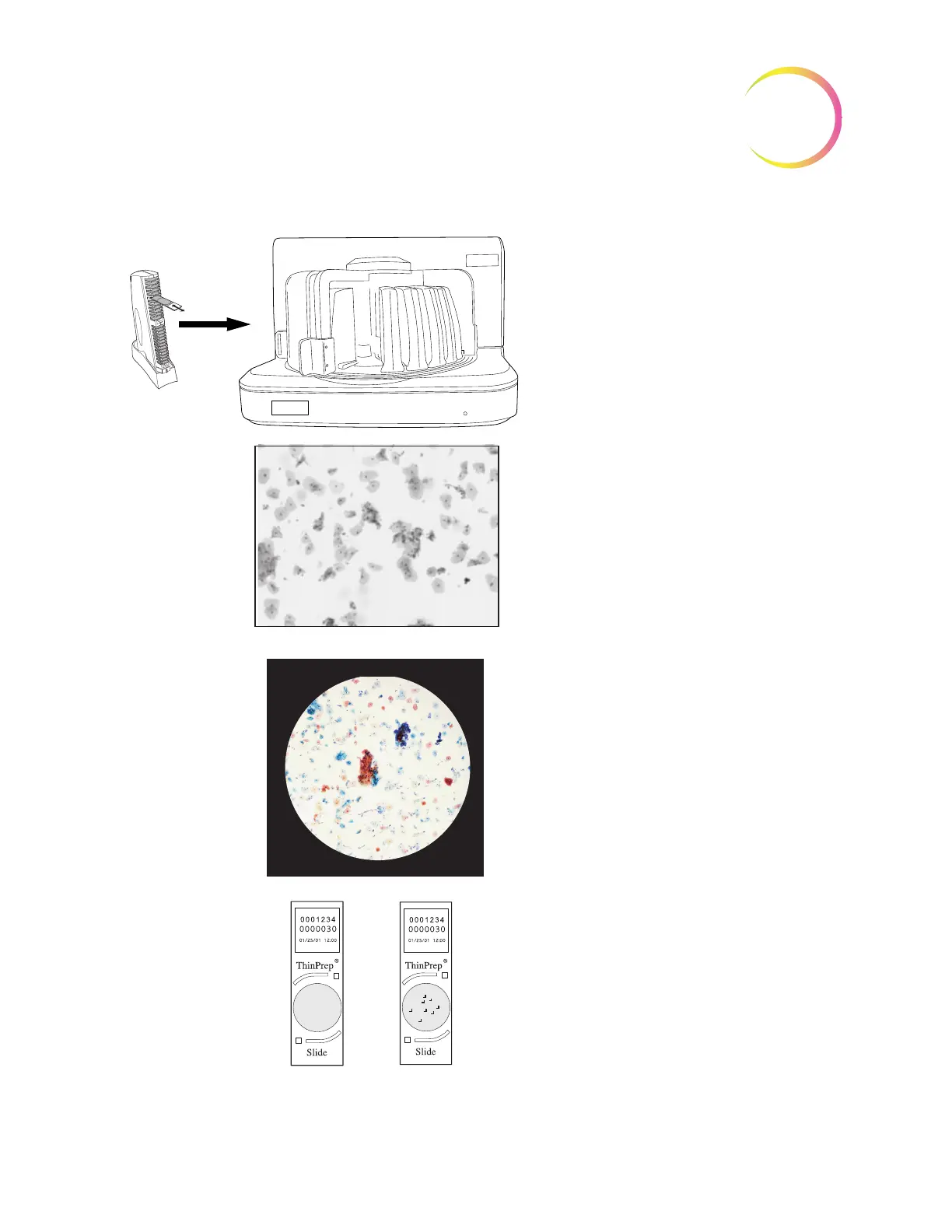
Prepared ThinPrep slides are
loaded into a slide cassette, which
is loaded into the Imaging Station
The slide imaging system scans the entire
cell spot. The system identifies objects of
interest found on the slide.
The coordinates of 22 objects of interest with
the highest integrated optical density will be
stored in the computer’s database.
The cell spot is
imaged
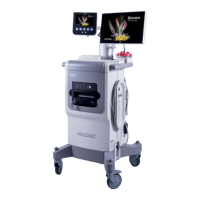
Slide review by the
Cytotechnologist
During Autolocate the system presents the 22
selected fields of view in geographic order to
the Cytotechnologist.
Suspect cells may be electronically marked by
the CT and a review of the entire cell spot is
enforced. The slide is physically marked.
At completion, the slide data is updated with the
location of any marked areas as well as infor-
mation on the review session.
Normal slide
Abnormal slides are reviewed by a
cytopathologist for interpretation and
diagnosis