5-6 Function and Performance Checking Method
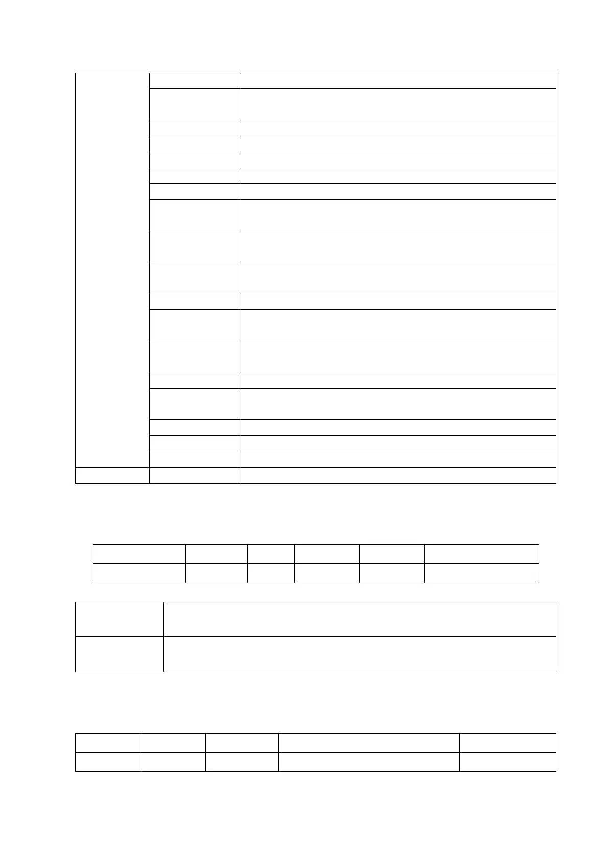
Changes the current probe frequency.
Turns on/off the colorize function; Selects among available
colorize maps.
Inverts the image vertically or horizontally.
Rotates an image at an angle of 90° each time.
Turns on/ off ExFOV function of probe to extend image range.
Merges images of two windows in Dual mode.
Adjusts contrast resolution of an image, compresses or expands
gray display range.
Selects among post processing map curves to optimize
grayscale images.
The function determines the quality and information of the
image.
Changes the number of focuses.
Increases image profile, so as to distinguish the image
boundary.
Optimizes the image by selecting acoustic speed according to
tissue characteristics.
Removes image noise to make details to be clearer.
Superimposes and averages images of different steer angles to
obtain image optimization.
Display or hide the width scale (horizontal scale).
Display different image effects of one probe.
Adjust gain of scan lines to increase the image lateral resolution.
Selects the acoustic power value.
2. M mode
In M mode scanning, the image parameter area in the upper left corner of the screen displays
the real-time parameter values as follows:
Parameters that can be adjusted to optimize the M Mode image are indicated in the following.
Gain, Depth,TGC,Focus Location
Speed, Display Format, Gray Map, Dynamic Range, Tint Map, M Soften, Edge
Enhance
3. Color mode
In Color mode scanning, the image parameter area in the upper left corner of the screen
displays the real-time parameter values as follows:
Pulse Repetition Frequency (PRF)
 Loading...


