EasyManuals Logo

Mindray DP-10 User Manual

Mindray DP-10
129 pages
To Next Page IconTo Next Page
To Previous Page IconTo Previous Page
Page #93 background imageLoading...
Page #93 background image
Transducers and Biopsy
12-23
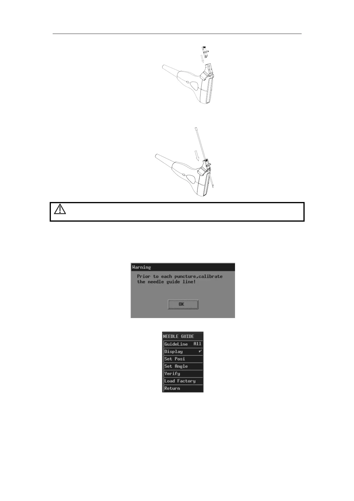
5) Insert a biopsy needle with the same specification as that of the guiding block into
the hole of the guiding block.
12.2.5 Entering Status of Guide Line Adjustment
1 When the B image is real-time, select [Puncture] in the [B MODE MENU] menu. The
following dialog box pops up.
2 Click [OK] in the dialog box. The [NEEDLE GUIDE] menu and guide lines appear.
Each needle-guided bracket has three guide lines at most.
When the image depth is between 2.16cm and 8cm, interval of two adjacent dots is
0.5cm;
When the image depth is between 8cm and 24.8cm, interval of two adjacent dots is
1cm.
CAUTION:
Ensure that all guide parts are seated properly prior to performing
a biopsy.

Table of Contents

Questions and Answers:

Question and Answer IconNeed help?

Do you have a question about the Mindray DP-10 and is the answer not in the manual?

Mindray DP-10 Specifications

General IconGeneral
Gray Scale256 levels
Weight5.5 kg
Imaging ModesB, B/B, 4B, M
Frequency2.0 to 10MHz (depending on the transducer)
Cine Loop256 frames
Power SupplyAC 100-240V, 50/60Hz
Image StorageBuilt-in storage, USB flash disk

Related product manuals