EasyManuals Logo

OCULUS Pentacam Interpretation Guide

OCULUS Pentacam
216 pages
To Next Page IconTo Next Page
To Previous Page IconTo Previous Page
Page #19 background imageLoading...
Page #19 background image
17
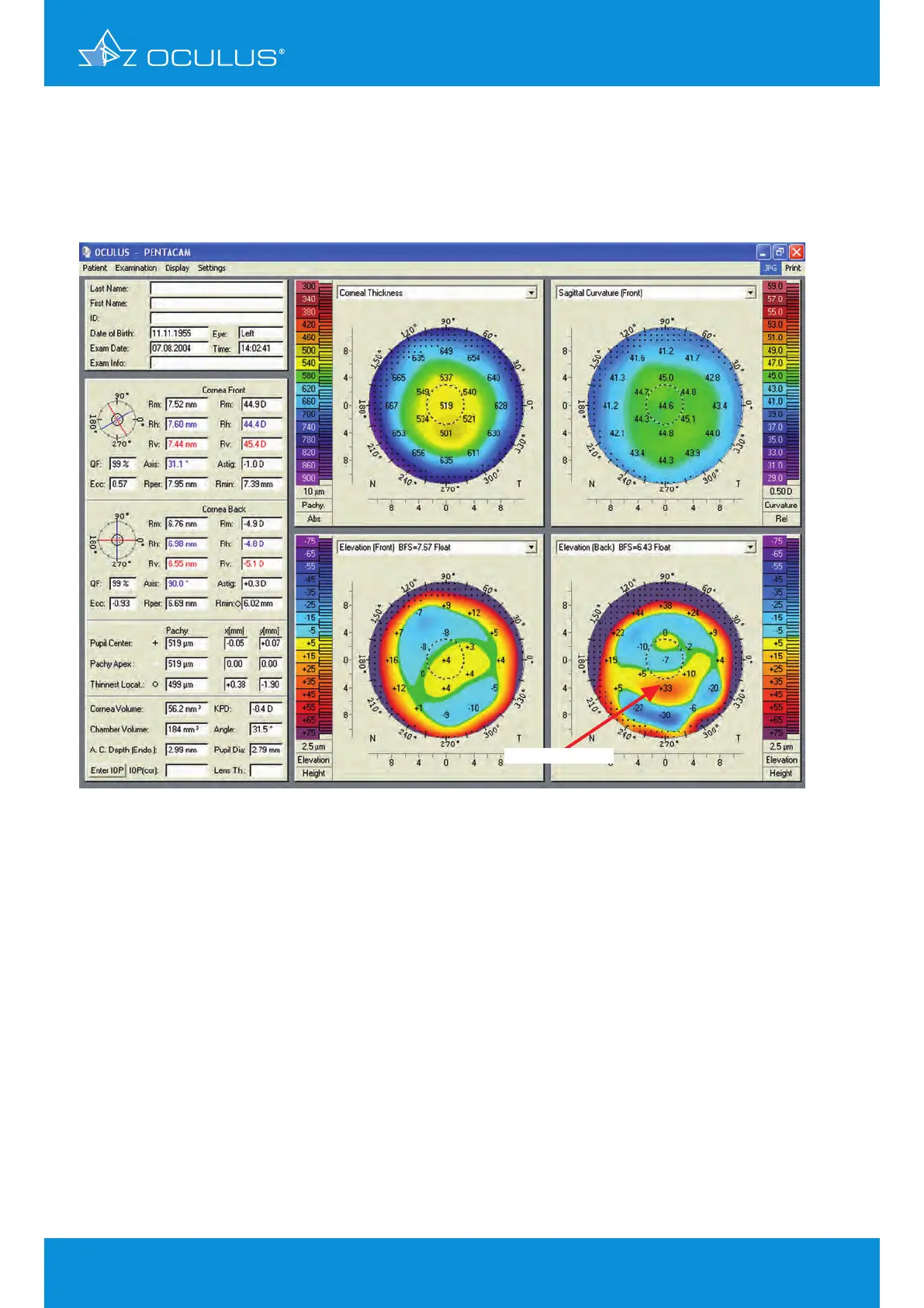
The left eye (Figure 10) indicates an inferior steepening, but a smooth anterior elevation map.
The reason for the thinning in the pachymetry map is the posterior elevation map, where there
are significant elevations of more than 30 μm. Note that the position of the thinning in the
pachymetry map and the highest spot on the elevation map are exactly at the same position.
This is an excellent example to document that topography or anterior elevation only does not
indicate keratoconus.
Figure 10: 4 Maps Selectable showing significant elevation in OS
significant elevation
5 Differences between Placido and
elevation-derived curvature maps

Table of Contents

Other manuals for OCULUS Pentacam

Questions and Answers:

Question and Answer IconNeed help?

Do you have a question about the OCULUS Pentacam and is the answer not in the manual?

OCULUS Pentacam Specifications

General IconGeneral
Measurement TimeLess than 2 seconds
Corneal CoverageUp to 12 mm
SoftwarePentacam Software
Data TransferUSB
Corneal TopographyYes
DisplayExternal monitor required
Measurement PrincipleScheimpflug imaging
CameraRotating Scheimpflug camera
Light SourceBlue LED
PachymetryYes
Anterior Chamber AnalysisYes
Imaging PrincipleScheimpflug imaging
FixationInternal
Power Supply100-240 V, 50/60 Hz
ResolutionHigh resolution
Lens Density MeasurementYes
DimensionsVaries depending on model and configuration
WeightVaries by model, check manufacturer specifications
Number of Images25 or 50

Related product manuals