117
16 INTACS
®
implantation
16 INTACS
®
implantation
16.1 Case 1: INTACS
®
implantation
by Prof. Michael W. Belin
A 27-year-old female was referred by her optometrist because of poor vision OD secondary to
keratoconus. Her BSCVA was 20/200 OD and with RGP over-refraction 20/30. The patient complained
of poor contact lens tolerance with less than 3 hours of daily wearing time. The patient was being
considered for intrastromal corneal ring segment implantation (ICRS, commonly referred to as
INTACS® in the US)
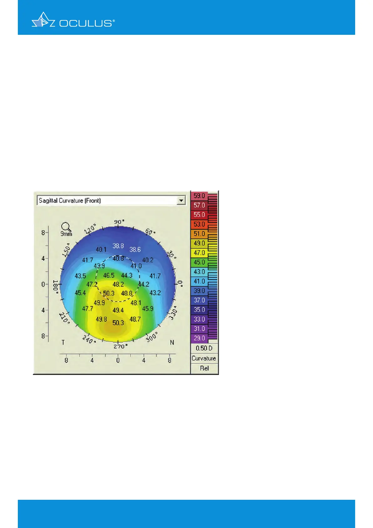
Anterior corneal curvature analysis showed significant inferior cone displacement and a maximum
steepness of > 50 D, with the steepest part of the cone well below the pupillary margin (Figure 144).
A presumptive diagnosis of pellucid marginal degeneration (PMD) was made, and the initial surgical
plan was to implant dissimilar INTACS® for PMD.
Surgical planning also included identifying the steep axis for the incision and looking at the
pachymetry over the incision location to determine the incision depth (Figure 145).
Surgical planning included:
implantation of 0.35 INTACS®
incision at axis 155°
incision depth 440 μm
Figure 144: Topography in a case of suspected PMD