EasyManuals Logo

OCULUS Pentacam Interpretation Guide

OCULUS Pentacam
216 pages
To Next Page IconTo Next Page
To Previous Page IconTo Previous Page
Page #170 background imageLoading...
Page #170 background image
168
23 Case reports from daily practice
23.5 Case 5: Incisional edema
by Prof. Renato Ambrósio Jr
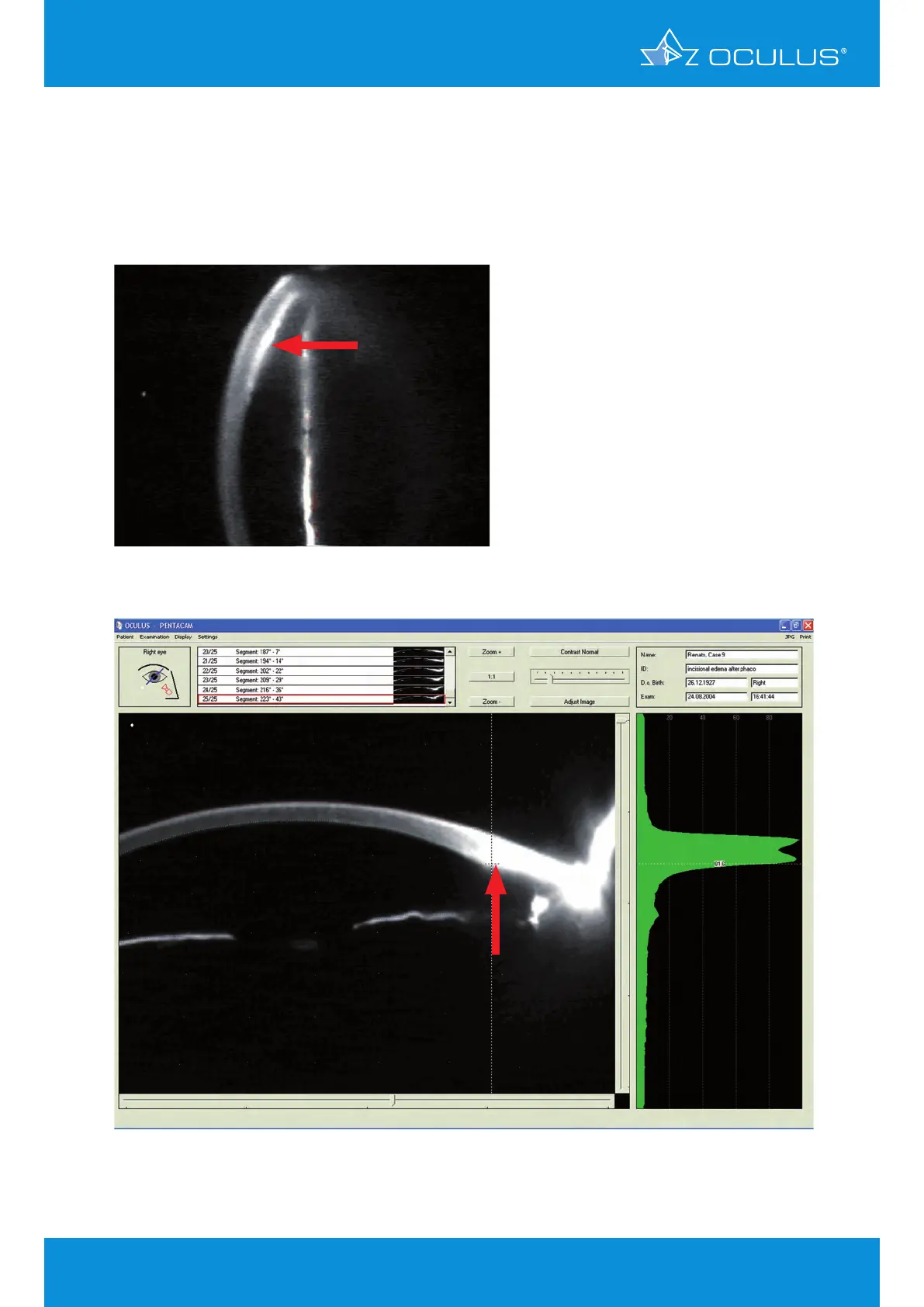
A 76-year-old female patient presented with incisional edema 12 months after phacoemulsification.
Endothelial morphology revealed large cells with pleomorphism and polymegathism. Central cell
count was 1.079 cells/mm².
Figure 202: Scheimpflug Image showing incisional edema
Figure 201: Slit lamp photo showing incisional edema

Table of Contents

Other manuals for OCULUS Pentacam

Questions and Answers:

Question and Answer IconNeed help?

Do you have a question about the OCULUS Pentacam and is the answer not in the manual?

OCULUS Pentacam Specifications

General IconGeneral
Measurement TimeLess than 2 seconds
Corneal CoverageUp to 12 mm
SoftwarePentacam Software
Data TransferUSB
Corneal TopographyYes
DisplayExternal monitor required
Measurement PrincipleScheimpflug imaging
CameraRotating Scheimpflug camera
Light SourceBlue LED
PachymetryYes
Anterior Chamber AnalysisYes
Imaging PrincipleScheimpflug imaging
FixationInternal
Power Supply100-240 V, 50/60 Hz
ResolutionHigh resolution
Lens Density MeasurementYes
DimensionsVaries depending on model and configuration
WeightVaries by model, check manufacturer specifications
Number of Images25 or 50

Related product manuals