102
12 Belin/Ambrósio Enhanced Ectasia Display
13 Locating the cone
13 Locating the cone
by Prof. Michael W. Belin
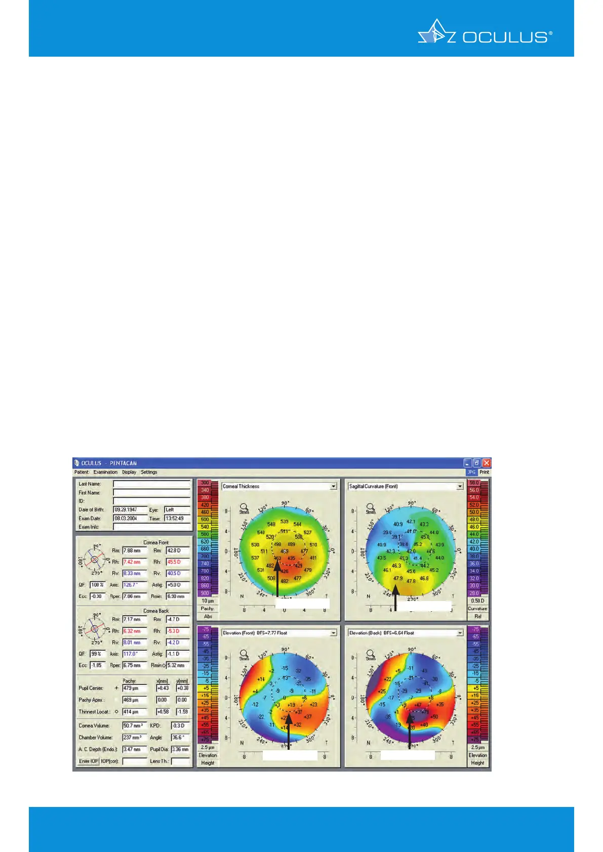
Most clinicians have characterized keratoconus based on the appearance on curvature maps. This
leads to inaccurate placement of the cone and a high incidence of supposed “pellucid marginal
degeneration”, which is a relatively rare occurrence. Elevation and pachymetry maps are more
reliable in locating the apex of the cone. The example below shows such a case.
Judging from the sagittal curvature map you would expect the cone between 6 and 7 o’clock. The
elevation maps of the anterior and posterior corneal surface show the rather position (Figure 122).
Refractive surgery screening commonly involves Placido disk based corneal topography and
central corneal thickness measurement by ultrasound [14,15,16]. At the time of its introduction
the ectasia risk score system, which is based on a topographic classification, proved to be an
improvement of the refractive surgery screening process [17,18]. However, some studies have
shown this scoring system to have drawbacks, revealing high false positive as well as high false
negative rates [19, 20, 21, 22, 23]. There are well defined risks for ectasia after LVC, and these
may be related to the presence of (typically mild) ectasia preoperatively, or a procedure that
determined important changes in corneal biomechanics [24]. From the viewpoint of these concepts
any cornea may evolve into ectasia if surgery or trauma weakens its biomechanical structure. This
can occur as a result of LASIK due to a thick flap or excess tissue ablation, or simply after a blunt
trauma. The likelihood of ectasia developing depends not only on the structural susceptibility of the
cornea but also on the impact of surgery [25, 26]. The process of screening for the risk of ectasia
developing must therefore do more than only detect mild forms of keratoconus or related diseases
[27, 28, 29].
Figure 122: 4 Maps Selectable with different representations suggesting different
cone locations
false position
rather position
rather position
rather position