104
14.1 Keratic precipitates
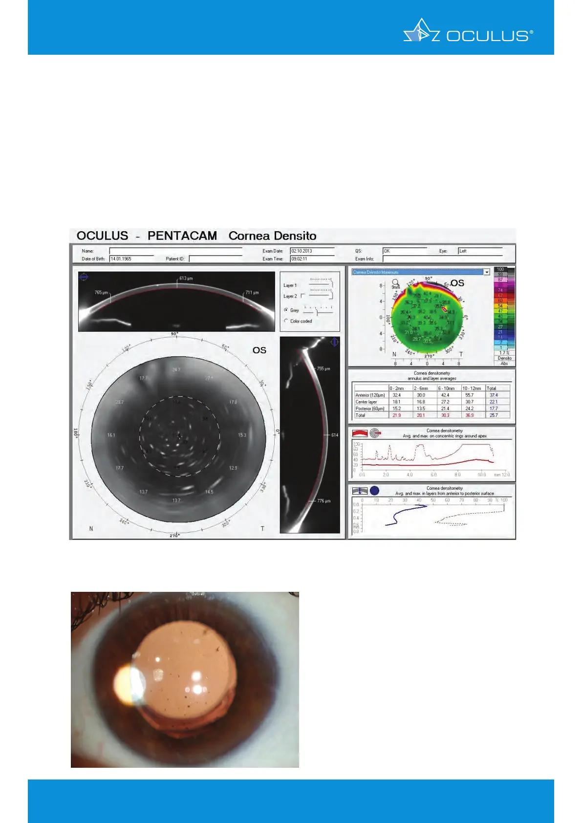
A 50-year-old patient presented with a history of granulomatous uveitis due to a toxoplasmosis
infection. At initial presentation he had numerous large keratic precipitates deposited on the
endothelial surface. In Figure 123 the large precipitates are prominent on the innermost layer of
the densitometry scan. Slit lamp photography was less successful in imaging the precipitates due to
the impossibility of targetingthe endothelial surface with retroillumination (Figure 124). Once the
patient had been started on a course of antibiotics and corticosteroids he showed significant clinical
improvement (Figure 125) and at his two week appointment there was no trace of the previous
keratic precipitates (Figure 126).
14 Corneal Optical Densitometry display
Figure 123: Corneal Optical Densitometry display showing a patient’s endothelial densi-
tometry at his initial presentation
Figure 124: Slit lamp photo of the same
eye at the patient’s initial
presentation