62
10.12 Keratoconus greater in OD than OS
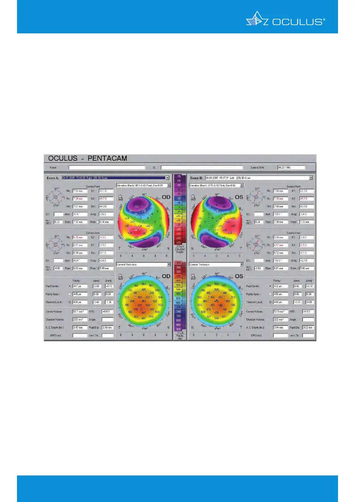
Look at the Show 2 Exams display of posterior elevation and pachymetry of OD and OS in this
patient with keratoconus (Figure 73). The right eye shows a significant posterior island (ectatic
area) associated with marked corneal thinning (430 μm) and significant inferior-temporal
displacement of the thinnest area towards the area of the abnormal posterior elevation. The left
eye shows a relatively normal posterior astigmatic pattern, but a distinctly abnormal pachymetry
distribution with marked inferior-temporal displacement and a thinnest reading of 440 μm. This
example shows the importance of looking at the pachymetry distribution, which may be the single
abnormal finding.
DIAGNOSIS - keratoconus greater in OD than OS
Figure 73: Show 2 Exams showing keratoconus greater in OD than OS
10 Screening for refractive surgery