58
10.9 Pellucid marginal degeneration
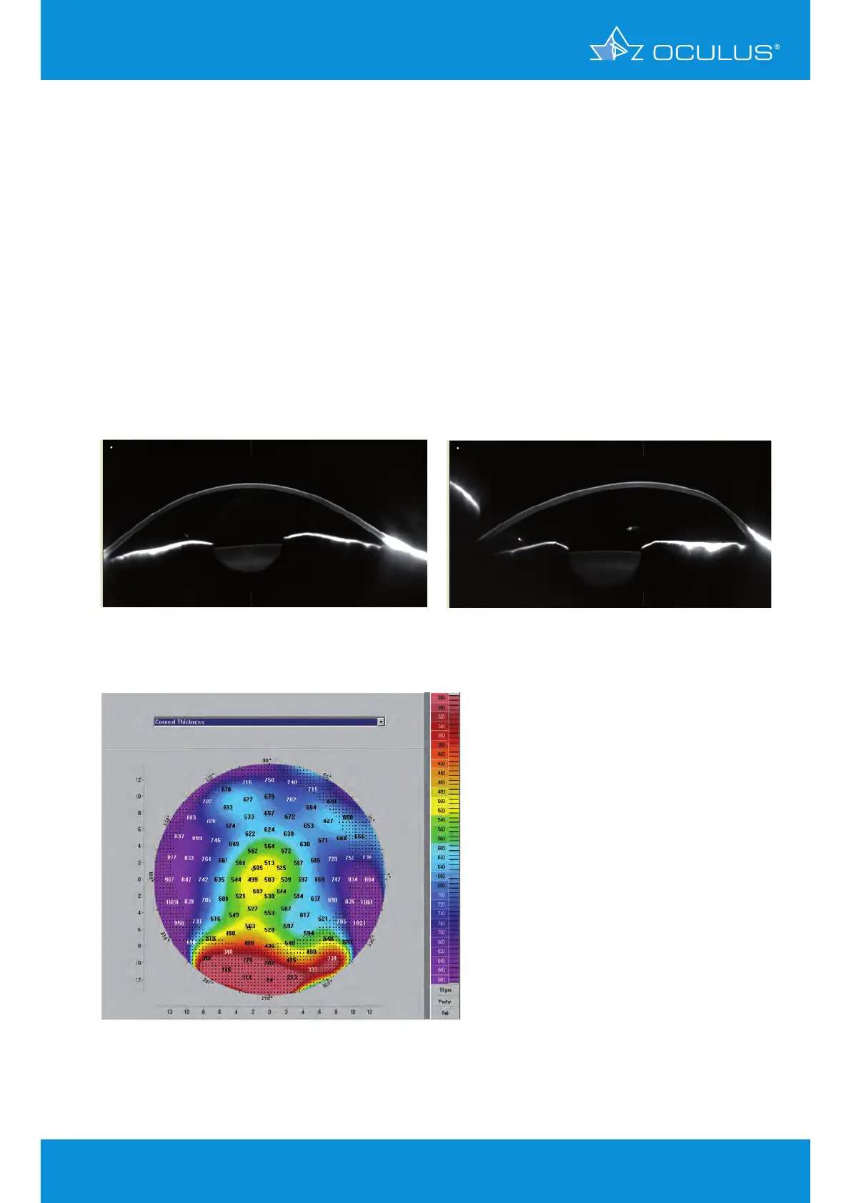
These are pictures of classic pellucid marginal degeneration (PMD). The pachymetry map
(Figure 69) shows the band of thinning located 1 - 2 mm from the inferior limbus. This is an area
that cannot be imaged on a Placido system, which is limited to imaging the central 9.0 mm.
The Scheimpflug images (Figure 67, Figure 89) show a relatively normal appearance when the
cornea is viewed through a horizontal cut and the pathognomonic appearance when viewed
through a vertical cross-section, revealing severe flattening over most of the cornea, an inferior
band of thinning and a sharp change in corneal contour over the area of thinning. PMD is one
of the most misdiagnosed conditions when the diagnosis is based on Placido imaging. A Placido
system cannot reach to the area of the pathology. Descriptive curvature terms such as "lobster
claw" pattern, etc. are fraught with problems and are associated with a very high false positive
rate.
DIAGNOSIS - pellucid marginal degeneration
Figure 69: Corneal thickness in a case of PMD
Figure 67: Scheimpflug image 180°
showing PMD
Figure 68: Scheimpflug image 90°
showing PMD
10 Screening for refractive surgery