100
12.6 Early ectasia with asymmetric keratoconus
by Prof. Renato Ambrósio Jr, Fernando Faria-Correia, MD,
Allan Luz, MD
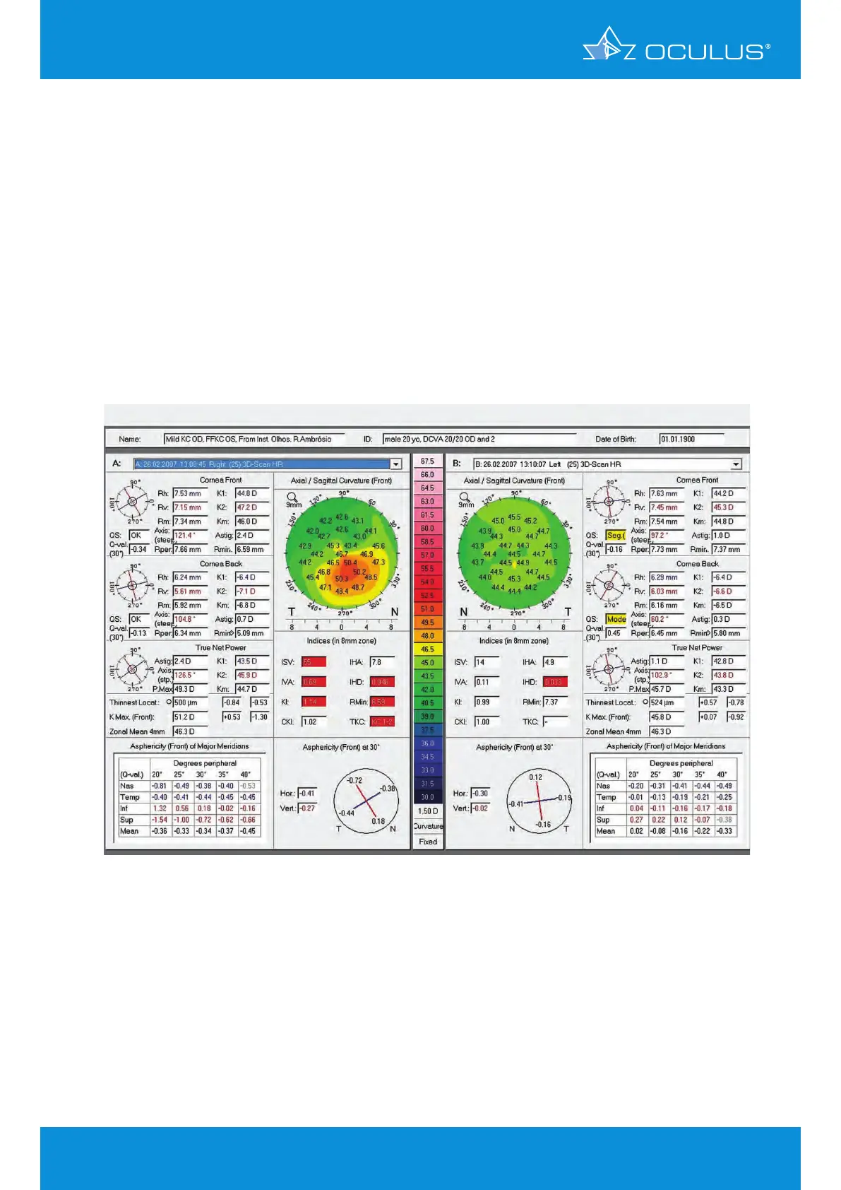
A 20-year-old male patient with asymmetric keratoconus presented with BCVA 20/20 in OD and
20/15 in OS. The data in OS show a relatively normal topography pattern, while those in OD reveal
mild keratoconus (Figure 119). Combining tomographic elevation and thickness data as it is done
in the Belin/Ambrósio Enhanced Ectasia Display improves the ability to detect ectatic disease [7,8].
The Ambrósio relational thickness maximum (ARTmax) [9] was 259 μm in both eyes (Figure 120,
Figure 121). The final deviation value D was 3.50 in OD and 1.77 in OS, which is consistent with
the clinical diagnosis. This is an example of a very mild forme fruste keratoconus in OS with still
relatively normal curvature topography and moderate tomographic changes [10,11,12,13].
Figure 119: Show 2 Exams anterior curvature sagittal map showing mild keratoconus
in OD and forme fruste keratoconus in OS
12 Belin/Ambrósio Enhanced Ectasia Display