EasyManuals Logo

OCULUS Pentacam Interpretation Guide

OCULUS Pentacam
216 pages
To Next Page IconTo Next Page
To Previous Page IconTo Previous Page
Page #114 background imageLoading...
Page #114 background image
112
15.1 Case 1: Corneal scar with RK incisions and cataract
This patient was referred with a central on-corneal scar with multiple RK incisions and cataract. As
a first step, he underwent scar peel with excimer laser myopic ablation to clear the cornea and make
it measurable. After IOL power calculation, he then underwent cataract surgery with a precisely
calculated lens implant and was brought to emmetropia and unaided 20/20 vision.
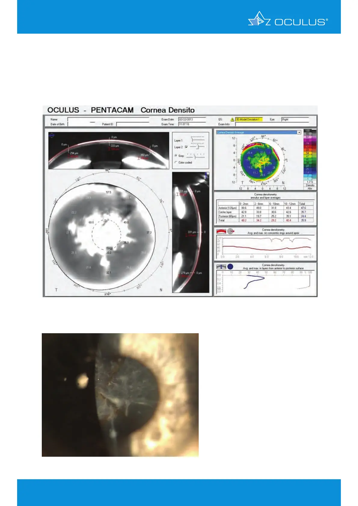
Figure 134: Corneal Optical Densitometry display showing a central corneal scar with
RK incisions and cataract
Figure 135: Slit lamp image of a central corneal scar with RK incisions and cataract
15 Using Pentacam
®
technology to evaluate corneal
scars, planning and documenting surgery outcomes

Table of Contents

Other manuals for OCULUS Pentacam

Questions and Answers:

Question and Answer IconNeed help?

Do you have a question about the OCULUS Pentacam and is the answer not in the manual?

OCULUS Pentacam Specifications

General IconGeneral
Measurement TimeLess than 2 seconds
Corneal CoverageUp to 12 mm
SoftwarePentacam Software
Data TransferUSB
Corneal TopographyYes
DisplayExternal monitor required
Measurement PrincipleScheimpflug imaging
CameraRotating Scheimpflug camera
Light SourceBlue LED
PachymetryYes
Anterior Chamber AnalysisYes
Imaging PrincipleScheimpflug imaging
FixationInternal
Power Supply100-240 V, 50/60 Hz
ResolutionHigh resolution
Lens Density MeasurementYes
DimensionsVaries depending on model and configuration
WeightVaries by model, check manufacturer specifications
Number of Images25 or 50

Related product manuals