18
5.2 Form fruste keratoconus?
A 47-year-old female presented for a second opinion. She had previously been told she was not a
candidate for refractive surgery and that she had “form fruste” keratoconus.
Her exam had revealed a BSCVA 20/20+ OD, and the slit lamp and external examination findings had
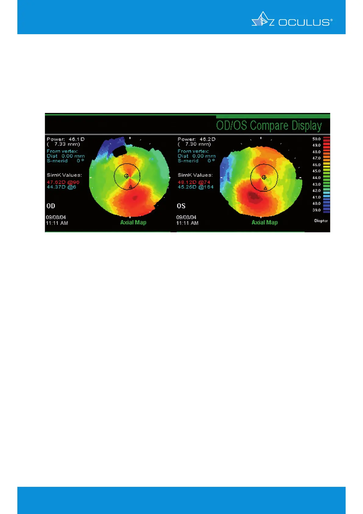
been WNL. However, Placido topography showed the following (Figure 11):
Pentacam® anterior segment analysis revealed normal pachymetry (normal distribution & central
thickness > 650 μm).
The anterior and posterior elevation revealed a slightly decentered apex. This had led to a “false
positive” inferior steepening on the curvature map. Custom LASIK was performed without incident
(Figure 12, Figure 13).
Note:
This case illustrates the limitations of curvature analysis in trying to analyze a shape abnormality.
Curvature is a reference-based measurement and in this case, inaccurately reflects shape
information. Elevation data are independent of axis or orientation and does not have the false
positive rates as curvature maps commonly do.
Figure 11: Placido based topography of OD and OS
5 Differences between Placido and
elevation-derived curvature maps