39
The ACA is 4º wider, and, although the ACD only deepened 0.09 mm centrally, the main difference
is evident in the periphery, where you can see changes ranging from 0.19 mm to 0.30 mm. This was
enough to increase the ACV from 64 to 92 mm³.
Comments
The Pentacam® is quite useful for measuring the ACA in narrow angle glaucoma, although this may
be difcult in 360º because of eyelid interference.
More consistent data can be obtained by measuring peripheral ACD and ACV.
The exam has been of great help also in educating the patient about this disease and making the
effect of the treatment evident to her.
9.3 Screening for narrow angles
by Dilraj S. Grewal, MD
9.3.1 Case 1
A 64-year-old Indian female patient presented for a routine eye exam. Her vision was 20/20 in
both eyes. She was found to have a shallow anterior chamber on slit lamp biomicroscopy (Figure
44). Gonioscopy showed Shaffer’s Grade 1 in all quadrants in both eyes. These findings were
confirmed by Scheimpflug images showing a shallow ACD of 1.80 mm in OD and 1.83 mm in OS.
The ACV was 64 mm
3
in both eyes. The ACA was 20.6 degrees in OD and 19.7 degrees in OS. The
ACD ratio was 0.5 in both eyes. Central corneal thickness was 557 μm in OD and 589 μm in OS
(Figure 45, Figure 46).
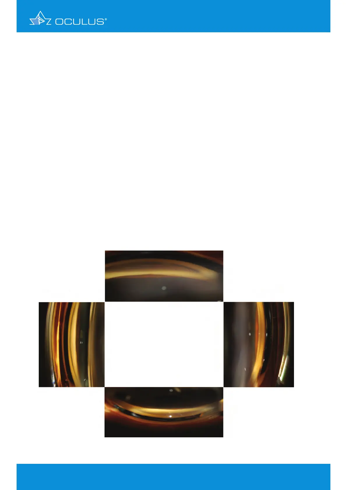
Figure 44: Slit lamp gonioscopy pictures showing a narrow angle in all four quadrants
9 Glaucoma