EasyManuals Logo

OCULUS Pentacam Interpretation Guide

OCULUS Pentacam
216 pages
To Next Page IconTo Next Page
To Previous Page IconTo Previous Page
Page #76 background imageLoading...
Page #76 background image
74
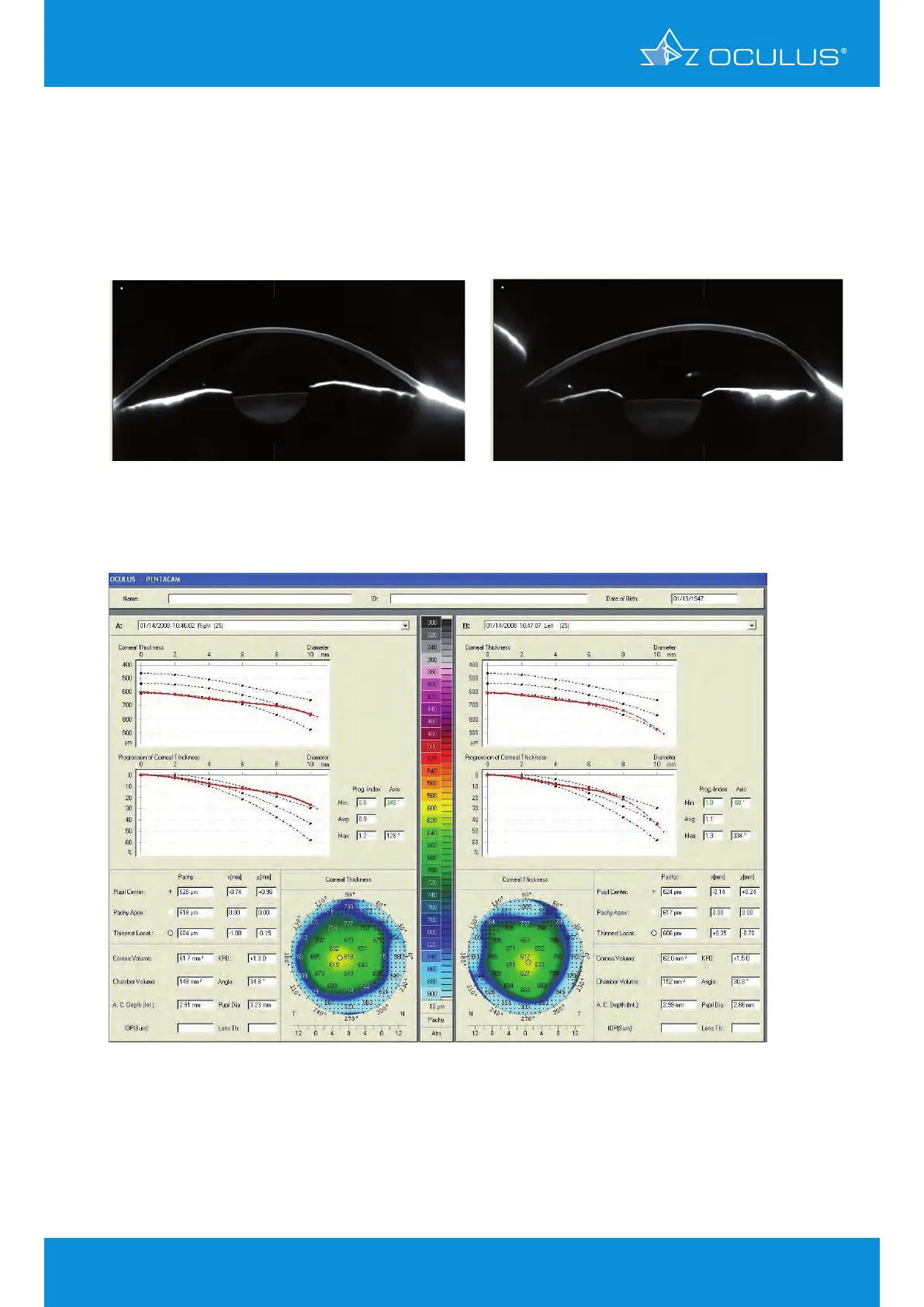
Figure 89: Show 2 Exams Pachymetric showing thick corneas with abnormal
corneal thickness progression in OD and OS
Figure 87: Scheimpflug image showing clear
cornea in OD with no peak in the
densitogram for the endothelium
Figure 88: Scheimpflug image showing clear
cornea in OS with no peak in the
densitogram for the endothelium
11.3 Case 2: Ocular hypertension
by Prof. Renato Ambrósio Jr, Marcela Q. Salomão, MD
The case below shows a patient with ocular hypertension. Please note the clear appearance of the
corneas in the Scheimpflug images (Figure 87, Figure 88) below as well as their thickness in the
Show 2 Exams display (Figure 89).
11 Corneal Thickness

Table of Contents

Other manuals for OCULUS Pentacam

Questions and Answers:

Question and Answer IconNeed help?

Do you have a question about the OCULUS Pentacam and is the answer not in the manual?

OCULUS Pentacam Specifications

General IconGeneral
Measurement TimeLess than 2 seconds
Corneal CoverageUp to 12 mm
SoftwarePentacam Software
Data TransferUSB
Corneal TopographyYes
DisplayExternal monitor required
Measurement PrincipleScheimpflug imaging
CameraRotating Scheimpflug camera
Light SourceBlue LED
PachymetryYes
Anterior Chamber AnalysisYes
Imaging PrincipleScheimpflug imaging
FixationInternal
Power Supply100-240 V, 50/60 Hz
ResolutionHigh resolution
Lens Density MeasurementYes
DimensionsVaries depending on model and configuration
WeightVaries by model, check manufacturer specifications
Number of Images25 or 50

Related product manuals