76
11.4 Case 3: Early Fuchs’ dystrophy with glaucoma
by Prof. Renato Ambrósio Jr, Marcela Q. Salomão, MD
This 60-year-old patient was referred to us for a second opinion on his diagnosis of normal tension
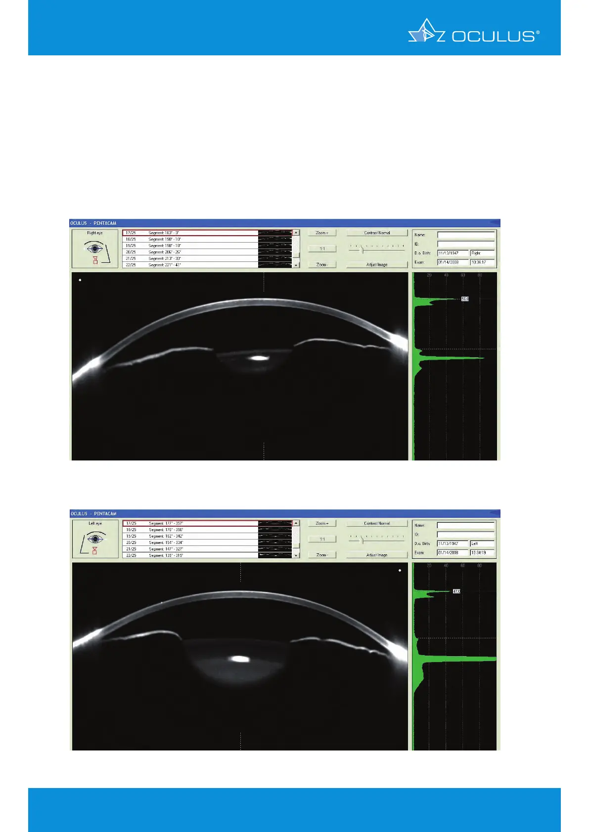
glaucoma, corneal disease and early cataract. The Scheimpflug images show higher scatter (less
clarity) and a second peak at the level of Descemet’s membrane and the endothelium (Camel’s sign) in
both eyes (Figure 91, Figure 92). This indicates a less transparent cornea. Both lenses can be seen to
lack clarity, even with non-dilated pupils. In both eyes corneal thickness is slightly thicker than usual
and the corneal thickness progression curve runs almost horizontally, indicating early oedema.
Figure 91: Scheimpflug Image showing a hazy cornea in OD
Figure 92: Scheimpflug Image showing a hazy cornea in OS
Figure 85, Scheimpflug image showing a case of Fuchs’ dystrophy in OS
11 Corneal Thickness