119
16 INTACS
®
implantation
16.2 Case 2: INTACS
®
after PRK
by Alain-Nicolas Gilg, MD
A 45-year-old female had had photorefractive keratectomy (PRK) in both eyes 7 years earlier.
Before the laser surgery her visual acuity had been
OD: sph -7.50 cyl -0.50 A 170°
OS: sph -6.75 cyl -1.00 A 10°
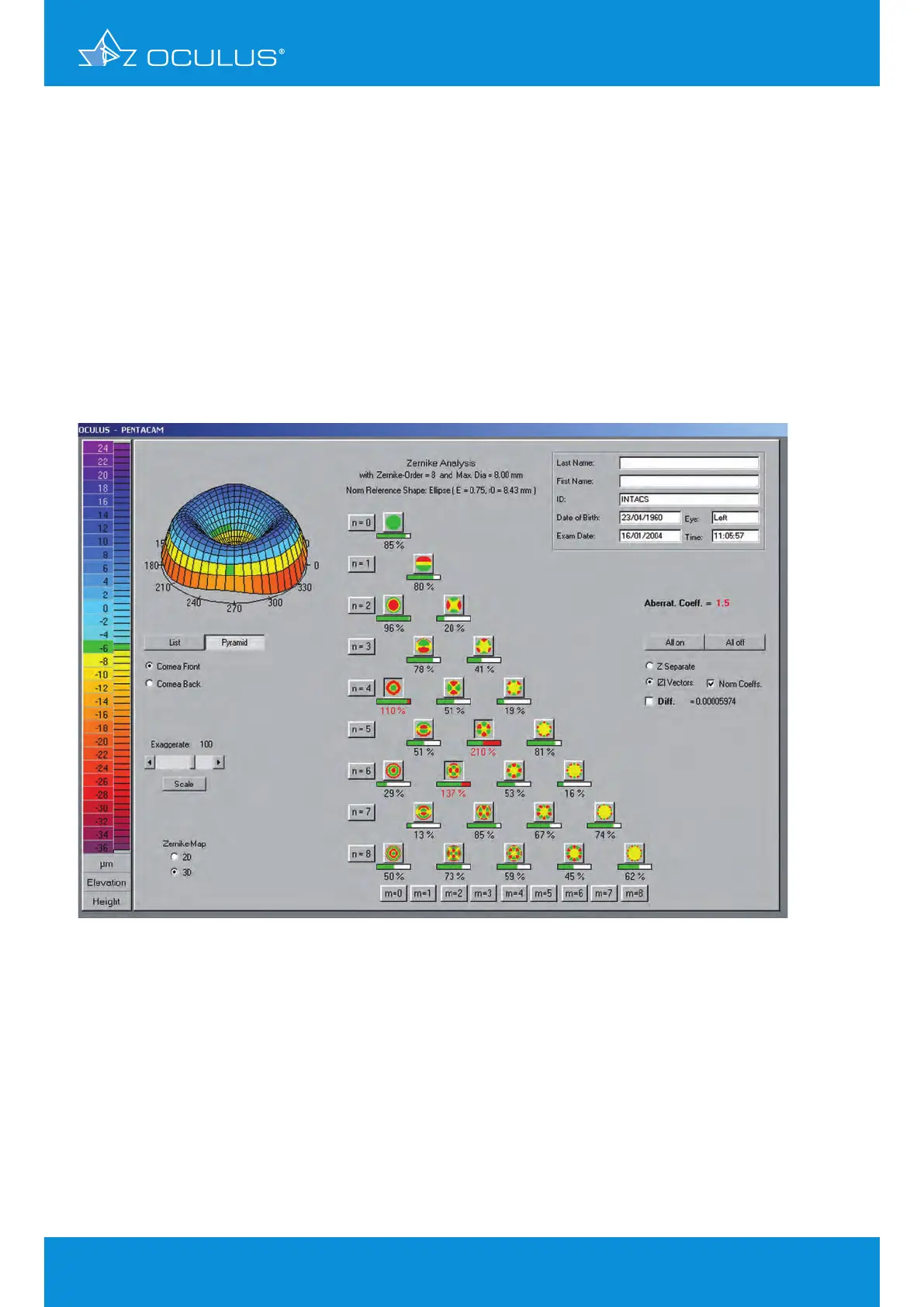
She was referred for blurred vision, photophobia, and poor intermediate VA. Zernike analysis
confirmed the functional disorders of her vision, showing them to be due to abnormal spherical and
high order aberrations (HOA), |Z|4° (spherical), |Z|5³ (trefoil 5
th
order) |Z|6² (astigmatism 6
th
order)
(Figure 147).
The keratoconus menu of the Pentacam® identifies this cornea as an oblate postoperative cornea.
Note the negative eccentricity and the abnormally high aberration coefficient due to the HOA
(Figure 148).
The pachymetry map shows a smooth progression with a thick area for the implantation of the
INTACS® in the 7 mm zone. This made her a good candidate for INTACS® implantation.
Before the implantation of corneal INTACS® her visual acuity was:
OD: sph -1.25, cyl -0.50 A 175° VA 20/25
OS: sph -1.50, cyl -0.50 A 55° VA 20/40
Figure 147: Zernike Analysis topography pre INTACS
®