118
16 INTACS
®
implantation
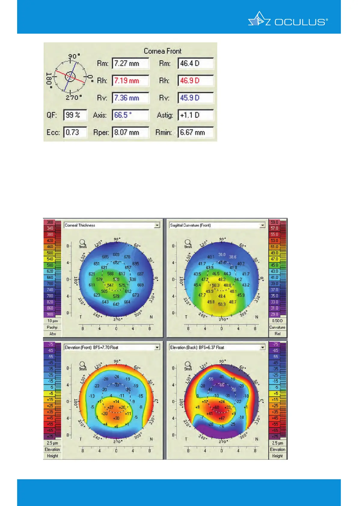
Then a complete Pentacam® anterior segment analysis was performed, revealing the shortcomings of
cone location and keratoconus classification based solely on anterior curvature.
Both the anterior and posterior elevation map, as well as the pachymetry map locates the cone just
at the inferior pupillary border, giving a picture typical of traditional keratoconus (Figure 145).
Figure 146: Part of 4 Maps Selectable showing a typical case of keratoconus
Figure 145: Keratometer values