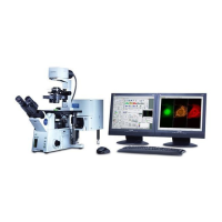
6. Optimizing Image Collection
The definition of “best” image will depend on the purpose(s) of the image and the nature of the
specimen. Sometimes the characteristics of a “best” image must be sacrificed in order to achieve more
important goals. A “pretty” picture is rarely suitable for quantitation or accurate representation of the
biology. Conversely, an image optimized for data is rarely pretty.
6.1 Qualities of an Optimal Confocal Image.
•
low noise
•
high specific (desired) signal
•
minimal bleedthrough between channels (spectral separation)
•
low background
•
high contrast
•
full dynamic range
•
absence of saturated pixels, at least in regions of importance
•
absence of pixels with intensity values of zero
•
absence of photobleaching or phototoxicity artifacts
•
spatial resolution
•
temporal resolution
These qualities are contradictory and compromises are required to achieve the “best” image from any
given specimen.
6.1.1 Overview of image capture properties
1. Try to keep ‘HV’ below 750 to minimize detector noise, or use averaging;
2. Minimize laser power levels to reduce bleedthrough and photobleaching;
3. Channel ‘Offset’ should be adjusted to remove blue pixels, usually ~5 to 7;
4. HV and laser power levels should avoid saturation (red mask);
5. Establish imaging conditions using monochrome display rather than color;
6. Determine how much resolution is required.
7. Collect some trial to test your parameters and possible changes.
6.2 Overview of basic image capture.
Several parameters must be balanced for optimal images including PMT settings, laser power, filter
selection, box size, zoom, choice of objective and dwell time.
There is no magic button on a confocal that will automatically provide the “best” image for you. Any
combination of settings that are appropriate for a given specimen and intended use of the images will
probably not be appropriate for a different specimen, different specimen preparation or different use of
the images. Therefore, it is important to understand how the confocal microscope works and
employ adequate control specimens.
6.2.1 Basic settings for confocal microscopy
1. Select dyes;
2. Find the specimen with the eyepieces with epi-fluorescence or transmitted light;
Olympus Fluoview-1000 User’s Guide
V.M. Bloedel Hearing Research Center, Core for Communication Research
Center on Human Development and Disability, Digital Microscopy Center
May 11, 2011 31
 Loading...