a. Image Acquisition Control Window>Click on transmitted light, Figure 4.1;
b. Image Acquisition Control Window>adjust brightness (TR Lamp) (click or scroll),
Figure 4.4;
6. Bring sample into focus;
7. Set Koehler illumination [optional, Section 7.3];
8. Center the region of interest in the field of view;
9. Turn off the transmitted light with the Image Acquisition Control Window, Figure 4.1;
10. Switch to fluorescent imaging.
Remove the DIC analyzer from the light path for optimal confocal images!
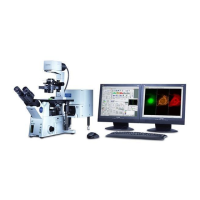
Figure 2. Enlarged view of Fluoview windows.
2.1 Menus
2.2 Acquisition settings
2.4 Microscope controller
2.3 Image acquisition
2.5 Dye list window
Olympus Fluoview-1000 User’s Guide
V.M. Bloedel Hearing Research Center, Core for Communication Research
Center on Human Development and Disability, Digital Microscopy Center
May 11, 2011 8
 Loading...