6 Acquiring Images
88 Basic User Manual
Line-Pos
SV-Size
s
0
5
10
15
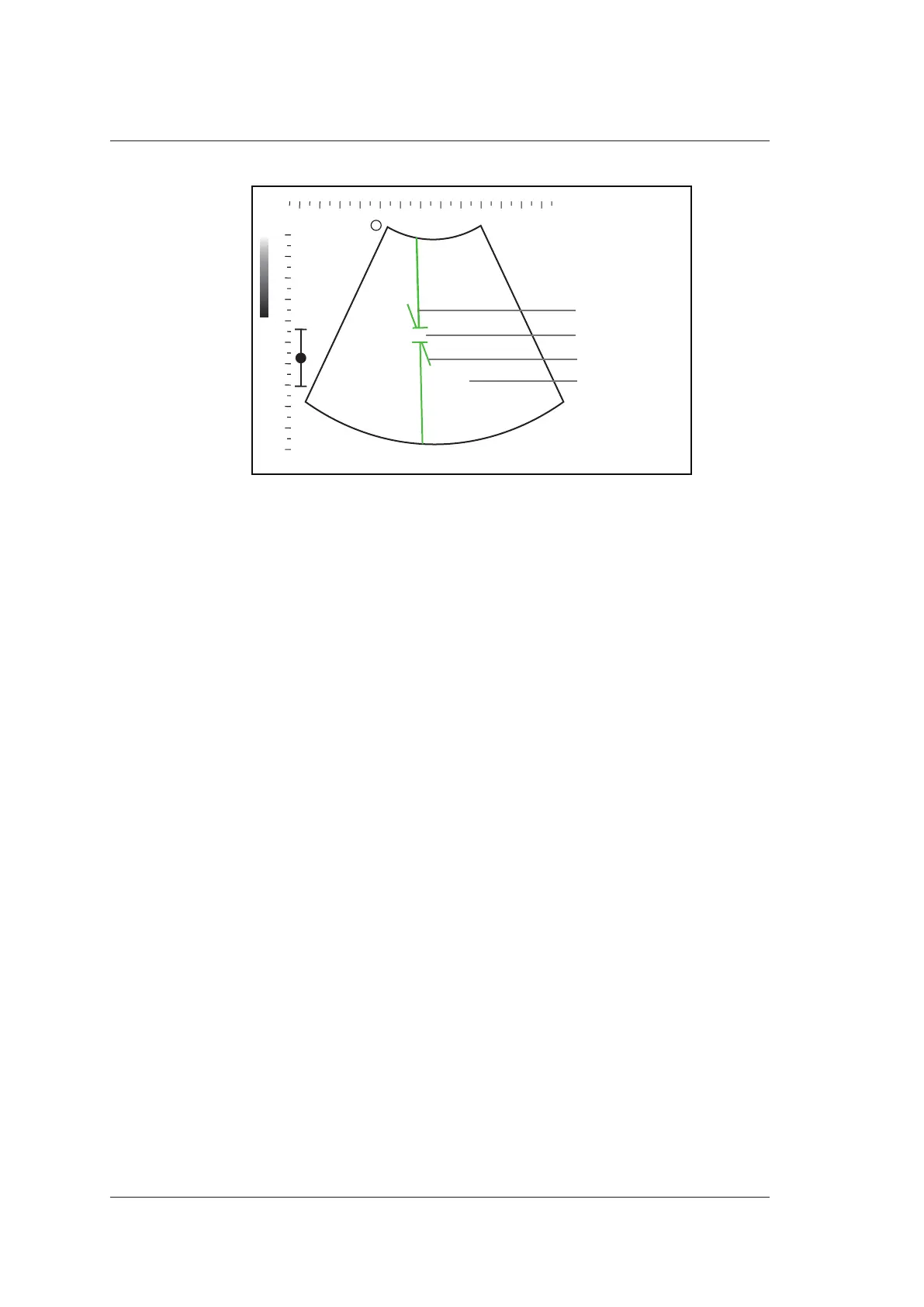
Spectral Doppler Line
Sample Volume Gate
Flow Cursor
2D Image
Figure 6-7 Inactivated B+PW Mode 1 Imaging Screen
−
The spectral Doppler line and the sample volume gate are used to locate the
qualitative analysis on the image.
−
The flow cursor needs to be adjusted parallel to the flow when measuring the
velocity.
3. Select Line-Pos by pressing the confirm key to adjust the position and angle of the
spectral Doppler line.
−
Position the sample volume gate on the spectral Doppler line by moving the
trackball up or down.
−
Adjust the angle of the spectral Doppler line by moving the trackball left or right.
4. Select SV-Size by pressing the confirm key and move the trackball to adjust the size
of the sample volume gate.
5. Rotate the Angle knob to adjust the angle of the flow cursor.
6. Press the Update key to activate the PW mode.
The PW spectrum is displayed at the lower part of the screen after being activated, as
shown in Figure 6-8.