Do you have a question about the Soredex DIGORA Optime and is the answer not in the manual?
Identifies and describes the components included with the DIGORA® Optime unit.
Details the necessary connections and components for setting up the DIGORA® Optime system.
Explains the unit's controls, buttons, and indicators for user interaction.
Provides step-by-step instructions on how to pack imaging plates correctly before use.
Guides users through the process of capturing and processing dental X-ray images.
Offers recommended exposure values for DC x-ray units to ensure optimal image quality.
Allows users to configure the DIGORA® Optime system to their clinical preferences.
Displays scanner type, firmware version, and unit serial number.
Explains image scanning settings like preview and dental chart.
Guides on marking teeth on the dental chart for image analysis.
Details Super (30 µm) and High (60 µm) resolution options.
Explains the function of noise filtering for smoother images.
Describes adding the unit serial number to new images.
Refers to installation for PC/LAN connection details.
Outlines the system's workflow options for image processing.
Explains automatic or manual start of image plate processing.
Notes that the system does not support touchless operation.
Describes options for plate ejection after processing.
Configures standby and shutdown timers for the unit.
Details settings for Comfort Occlusal™™ imaging.
Mentions FMS template feature availability for US users.
Introduces protective covers and hygiene bags for imaging plates.
Describes available imaging plate sizes and types.
Explains the function of the storage box for plate protection.
Recommends holders for accurate patient positioning and image quality.
Details the start-up kit for occlusal imaging.
Mentions microfibre cloth for dry cleaning.
Describes the chemical composition of cleaning wipes.
Provides guidelines for handling and caring for imaging plates.
Details the process and approved agents for cleaning imaging plates.
Explains what an imaging plate is and its benefits over film.
Covers protective covers and hygiene bags for plate protection.
Describes the steps involved in processing an imaging plate.
Discusses the effect of background radiation on imaging plates.
Explains the impact of ambient and UV light on imaging plates.
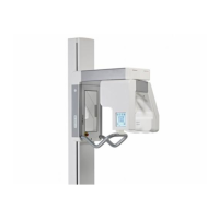
Guides on optimal placement of the unit for operation and maintenance.
Specifies placement guidelines for PC/network switch relative to the patient.
Details the procedure for connecting the unit to a PC or network.
Warns against connecting unauthorized external devices.
Explains common image errors and their causes.
Details issues arising from incorrect use of accessories and plates.
Covers errors related to incorrect X-ray settings and exposure.
Discusses issues related to imaging plate wear and tear.
Lists and explains system error codes and messages.
Outlines procedures for maintaining optimal system performance.
Provides safety precautions for cleaning and disinfecting the unit.
Lists approved cleaning agents and methods for the unit.
Details disinfection procedures and precautions for the unit.
States that the unit requires no user maintenance.
Directs repairs to authorized service personnel only.
Advises on proper disposal of the unit and accessories.
Lists device name, model, intended use, manufacturer, and standards.
Details PC, network, and software requirements for the system.
Provides specifications for different imaging plate sizes.
Lists specifications for hygiene bags.
Presents EMC emission and immunity guidance and tables.
Explains common symbols used on the unit and in documentation.
Shows the unit's main label with key identification information.
Lists critical warnings and precautions for safe operation.
| Resolution | Up to 20 lp/mm |
|---|---|
| Plate Sizes | 0, 1, 2, 3, 4 |
| Theoretical Resolution | 20 lp/mm |
| Software | DIGORA for Windows |
| Connectivity | Ethernet |
| Grayscale | 16 bits (65536 shades of gray) |
| Scanning Time | Approx. 10 seconds |