36
Publicaon 5423-020-REV 1.0 • www.btxonline.com
DNA Delivery into Cells
Using Electroporaon
General Electroporaon Discussion
Electroporaon is the use of a transmembrane electric eld pulse
to induce microscopic pathways (pores) in a biomembrane. Their
presence allows molecules, ions, and water to pass from one side
of the membrane to the other. When the electric eld is applied,
the ions inside and outside the cell membrane migrate. As the
charge builds up on either side of the membrane the membrane
weakens and the pathways form perming material outside
of the cell to enter. If the electric eld is promptly removed
the pathways close and the membrane reseals. If the electric
eld duraon is too long the pathways increase and the cell is
killed. Ecient electroporaon depends on proper selecon of
electric eld waveforms. The electropores are located primarily
on the membrane areas which are closest to the electrodes. The
pathways form in about a microsecond and seal in seconds to
minutes. The duraon of the electric eld is tens of microseconds
to tens of milliseconds.
The use of electroporaon was described by Neumann in the early
1980’s. The roune use of electroporaon became very popular
with researchers through the 1980’s because it was found to be a
praccal way to place drugs, or other molecules into cells. In the
late 1980’s, sciensts began to use electroporaon for applicaons
in mul-cellular ssue.
In the early 1990’s Lluis Mir of the Instute Gustave-Roussy was
the rst to use electroporaon in a human trial to treat external
tumors.
Research has shown that the inducon of pathways is aected by
three major factors. First, cell-to-cell biological variability causes
some cells to be more sensive to electroporaon than other
cells. Second, for pathways to be induced, the product of the
pulse amplitude and the pulse duraon has to be above a lower
limit threshold. Third, the number of pathways and eecve
pathway diameter increases with the product of “amplitude” and
“duraon.” Although other factors are involved, this threshold
is now understood to be largely dependent on a fourth factor,
the reciprocal of cell size. If the upper limit threshold is reached,
pore diameter and total pore area are too large for the cell
to repair by any spontaneous or biological process, and the
result is irreversible damage to the cell or cell lysis. Because
the mechanism of electroporaon is not well understood,
the development of protocols for a parcular applicaon has
usually been achieved empirically, by adjusng pulse parameters
(amplitude, duraon, number, and inter-pulse interval).
Research shows that certain experimental condions and
parameters of electrical pulses may be capable of causing many
more molecules to move per unit me than simple diusion.
There is also good evidence (Sukharev et al., 1992) that DNA
movement is in the opposite direcon.
An addional important consideraon when the voltage pulse is
applied to the cells and medium is that the amount of current that
ows is dependent on the conducvity of the material in which
the cells are located. Some material is quite conducve and severe
heang will occur if the pulse duraon is too long. Therefore long
duraon elds will kill cells by destroying the membrane and
heang.
The electric eld in which the cells are located is produced by two
system components. The rst is the voltage waveform generator
and the second is the electrode which converts the voltage into
the electric eld.
As the charge accumulates at the membrane, which is a
capacitance, the voltage across the membrane increases:
voltage = capacitance charge
As charge accumulates at the membrane, the voltage across
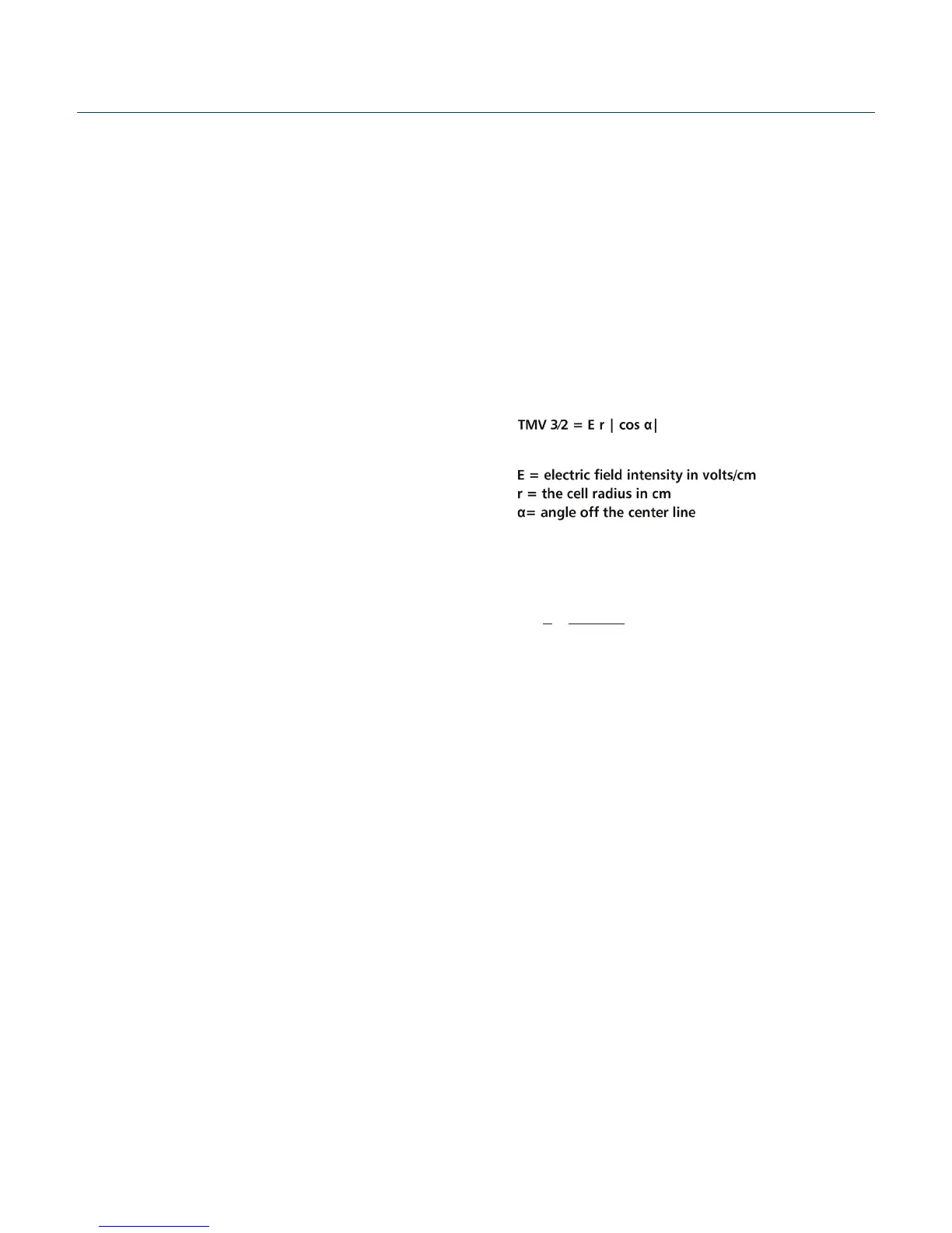
the membrane increases. Neumann et al. (1989) described the
equaon that relates the transmembrane voltage (TMV) to electric
eld intensity:
where:
Pores in the membrane will begin to form as the voltage increases
from its quiescent value of a few tenths of a volt to more than 0.5
volts. To produce a TMV of 1 volt across the membrane of a cell
with 7 μm radius, the required electric eld intensity is:
The number of pores and eecve pore diameter increase as
the product of pulse amplitude and duraon increase. At the
upper limit threshold, pore diameter and total pore area become
too large for the cell to repair by any spontaneous or biological
process. The result is irreversible damage to the cell or cell lysis.
Another important point to consider is the generaon of heat
during electroporaon. Heat producon is directly related to
current intensity which is, in turn, dependent on the conducvity
of the material through which the electric eld is applied.
Standard saline soluons such as PBS and many ssue culture
media are highly conducve and thus will generate considerable
amounts of heat when used in cell electroporaon. Excessive
heang can be detrimental to cell viability. The eects of heang
can be reduced by using a low conducvity medium such as BTX’s
Cytoporaon Medium to resuspend cells prior to electroporaon.
Although electroporaon is an eecve method for introducing
macromolecules onto cells, the biological mechanisms by which
cells become electroporated are not completely understood.
Therefore, the development of specic protocols for parcular
applicaons is usually achieved by empirical adjustment of
pulse parameters (i.e. amplitude, duraon, pulse number, and
interpulse interval).
E= =
2
3
950 volts/cm
1
7 x 10
-4
*
General Optimization Guide for Electroporation