5 Operating procedures
42 GE Healthcare 86500-IMG rev 3
5.10 SPECIAL EXPOSURES
5.10.1 Magnification studies
By placing the breast at a distance from the film causes the image to be
magnified. This increases the size of the smallest objects (micro-
calcifications) above the grain size of the film-screen combination thus
improving the image quality.
WARNING!
As source to skin distance is decreased when using a magnification
tunnel, avoid unnecessary magnification examinations or keep the focal
spot to the skin distances as large as possible in order to keep the skin
dose as low as possible. Do not perform screening with a magnification
tunnel!
The air gap between the breast table and the cassette reduces scattered
radiation and further improves image contrast. A small focal spot is
essential for good magnification image quality and therefore a true
0.1mm focus is needed. The Diamond X-ray tube features a Gaussian
0.1mm focal spot and during magnification studies this is automatically
selected.
Although the object may be visualized well, this is offset by the fact that
only a small area of the breast can be imaged at a time.
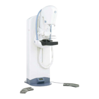
For magnification studies the Diamond uses the MultiChoice
magnification tunnel. One tunnel is used for all magnification factors 1.6x,
1.8x and 2.0x. The factor is a user preference and can be changed by the
user. The film format in magnification studies is always 18x24cm.
Magnification studies can be performed by applying compression to the
whole area of the breast, which is all imaged or by applying compression
only to a specific area, called spot compression. In whole field imaging, a
larger area can be studied on one image, for example, when it is not
known where the area of interest lies in the breast.
5.10.2 Magnification procedure
Remove the face shield and install
the magnification tunnel. The
magnification tunnel is inserted
and removed the same way as
the cassette holders. During
insertion of the magnification
tunnel, make sure that you hear
two clicks, one on each side. This
means that the tunnel is locked
and stable. If the tunnel is not
locked correct, it will slide down
during compression .
Follow the general patient
positioning procedure. The size of the top of the magnification tunnel
corresponds to the area which can be magnified onto the 18 x 24cm film
size.
FOR TRAINING PURPOSES ONLY!
NOTE: Once downloaded, this document is UNCONTROLLED, and therefore may not be the latest revision. Always confirm revision status against a validated source (ie CDL).