1111
Technical Description
4. Technical Description
4.1 The Doppler Principle
The Dopplex Range all use a technique based on the Doppler principle for non-invasively
monitoring movement within the body.
The Doppler principle states that if a signal is transmitted at a fixed frequency and is reflected
by a moving body, the frequency of the received signal will be shifted. An increase in
frequency results if the reflector is moving towards the transmitter/receiver, and a decrease
results if moving away from the transmitter/receiver. The amount of frequency shift is
proportional to the velocity of the reflector relative to the transmitter/receiver.
In the Dopplex range, a fixed frequency ultrasonic signal is transmitted from the probe into the
body. This is reflected from, for example, moving blood cells. The signal is reflected from
these cells and is received by the probe. Due to the movement of the blood cells, a frequency
shift results, which is proportional to the blood flow velocity. The Doppler shift is also affected
by the angle between the probe and the direction of flow. The Doppler shift is greatest when
the flow is directly towards, or away from, the probe.
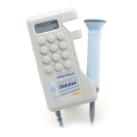
4.2 Doppler Audio Processing
The pocket Dopplex probe contains a transmitter and receiver. In use, the probe sends out a
continuous ultrasonic signal (carrier), generated by the piezo-ceramic transmitter crystal, in the
frequency range 2 to 10 MHz (depending on probe).
This signal is scattered by blood cells or any other "interface" such as skin, muscle layers,
organs, walls of vessels etc. A small proportion of the scattered signal will be reflected back
and detected by the receiver.
By demodulating the received signal (removing the high frequency carrier) the Doppler shifted
component (i.e. the difference between the transmitted and received signals) can be produced.
With typical target velocities found in the human body, this Doppler shift signal falls within the
audio frequency range. It can therefore simply be amplified and heard through a loudspeaker.
It is important to remember that the sound you hear is an artificial sound, the frequency (pitch)
of which is proportional to the velocity of the moving target. It is not the real sound made by
blood rushing through an artery or vein, or movement of the fetal heart.
4.3 Fetal Heart Rate Processing, (FD2-P, MD2-P, FD1-P, FD3-P)
In addition to providing this Doppler sound, the circuitry in the FHR signal conditioning section
generates an amplitude envelope of the Doppler audio signal. Using auto-correlation, this
signal is further processed by the microcontroller to calculate FHR.
4.4 Bi-directional Signal Processing (MD2-P, SD2-P, RD2)
To achieve bi-directional flow indication, the Doppler signal must be further processed to
separate forward and reverse components.
Components of the Doppler signal produced by positive frequency shift represent flow towards
the probe, referred to as forward flow. Components of the Doppler signal produced by negative
frequency shift represent flow away from the probe, referred to as reverse flow.
The circuitry achieves this separation in the vascular signal processing section producing two
frequency envelopes using zero crosser techniques. This signal is presented at the waveform
output (MD2-P, RD2).
The microcontroller also receives these bi-directional signals and displays them on the LCD
bargraph to give a visual indication of blood flow velocity and direction.
 Loading...