7-2 Physiological Signal
9. Display effect of respiratory curve depends on the patient
breathing status. While a very slow or smooth breathing may
lead to an inapparent respiratory curve, breathing in a large
amplitude may cause an incomplete display of the respiratory
curve.
10. Display effect is linked to the connected parts of the body.
Generally, signals by connecting to limbs are stronger than by
connecting to the chest.
When abnormality is detected in physio trace, please check if ECG leads are
properly connected with the system.
7.1 ECG
7.1.1 ECG Operation Basic Procedures
1. Connect the device.
Turn off the power supply of the system, and connect the ECG cable to the corresponding
interface on the physiological module.
Turn on the power supply of the system.
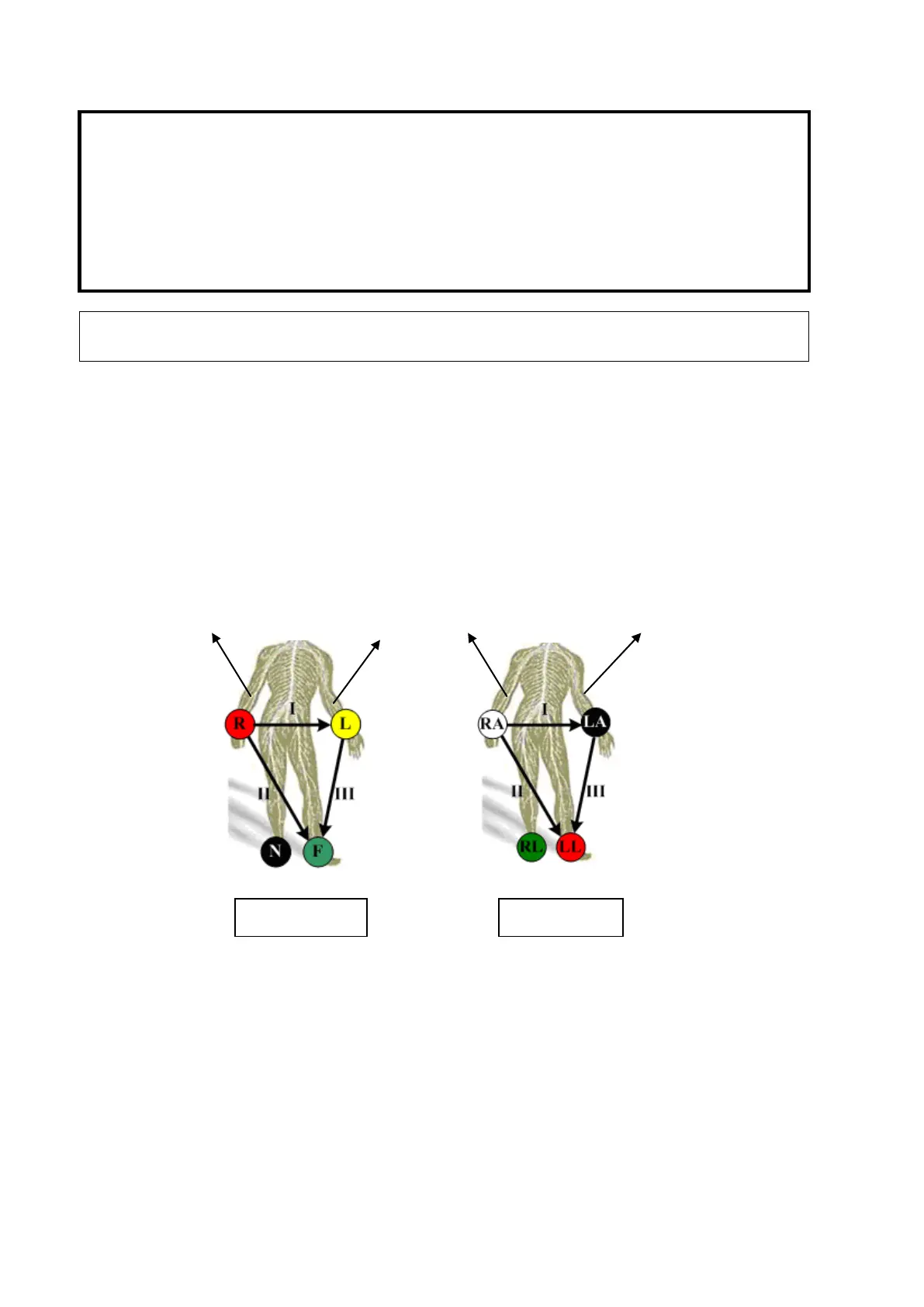
Place the ECG electrodes on the patient’s body (as shown in the following figures).
2. Press <Physio> key on the control panel to enter physio operation status.
3. Switch the imaging modes and display formats, adjusting the parameters to get an optimized
image.
4. Parameter adjustment: On the Physio page, you can adjust [Speed], [ECG Gain], [Position]
and [Invert].
5. Trigger:
Select the trigger mode, or switch on [Real & Trig], set the trigger time.
6. Freeze the images and review the images.
7. Press <Physio> to exit ECG mode, and remove ECG electrodes from the patient.