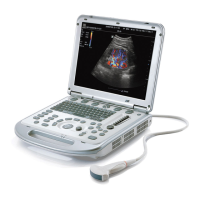
14
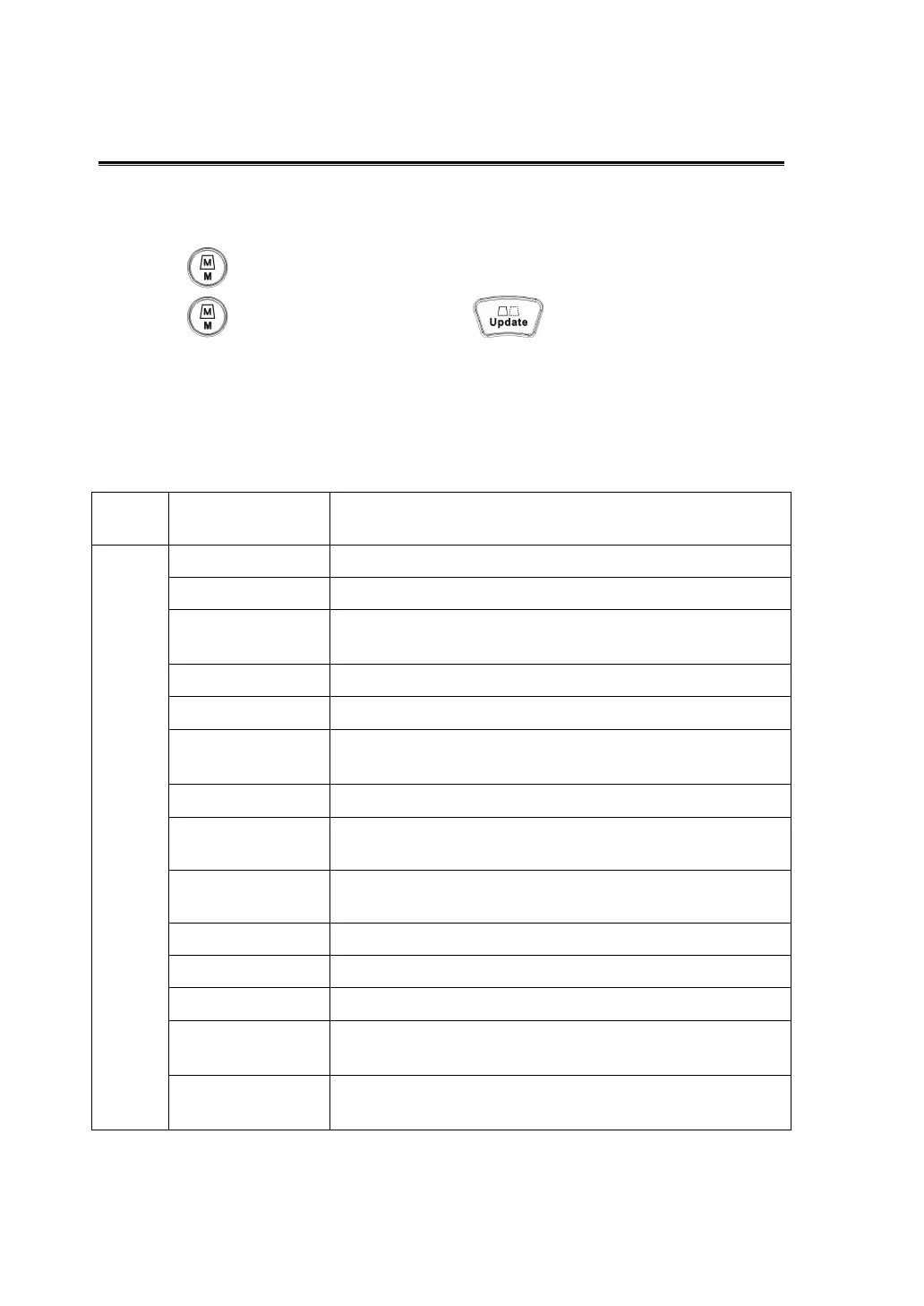
10 Image Optimization: M
1.
Select a high-quality image during B mode scanning, and adjust to place the area
of interest in the center of the B mode image.
2.
Press on the control panel, and roll the trackball to adjust the sampling line.
3.
Press on the control panel again or to enter M mode, then you
can observe the tissue motion along with anatomical images of B mode. During
the scanning process, you can also adjust the sampling line accordingly when
necessary.
4.
Adjust the image parameters to obtain optimized images.
5.
Perform other operations (e.g. measurement and calculation) if necessary.
Control
Panel
Gain Changes the M mode gain---- rotate <iTouch> knob.
Menu
Time Mark Turns on/ off the time mark display in M mode image.
Speed Increases or decreases the M mode sweep speed.
Display Format
Selects the display format of M mode image with B
mode image.
Frequency Changes the current probe frequency.
A. power
Selects the acoustic power value.
Gray Map
Selects among post processing map curves to optimize
grayscale images.
Focus Position
Changes the focus number.
Dynamic Range Adjusts contrast resolution of an image, compresses or
expands gray display range.
IP (Image
Processing)
Selects among groups of image combination
parameters to optimize the image.
Colorize Turns on/off the colorize map display.
Colorize Map Selects among available colorize maps.
M Soften
Rejects noise to make images clear.
Post Process
Gray correction for an image to obtain optimum map,
including curve,
γ
, Gray rejection.
Edge Enhance
Increases image profile, so as to distinguish image
boundary.