154
22 Phakic IOL implantation
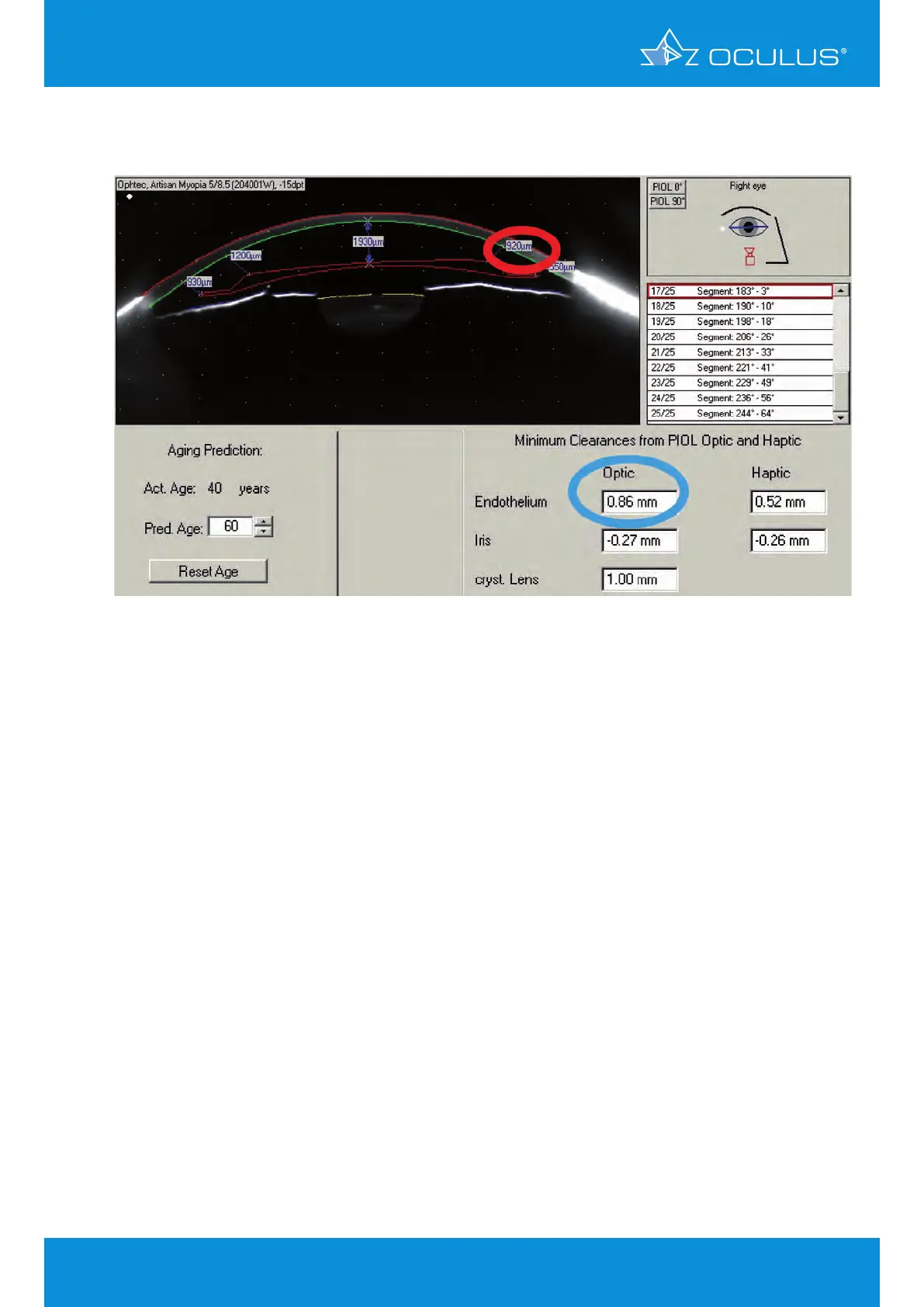
The image below shows the simulated pIOL position 20 years after implantation.
The horizontal Scheimpflug image shows a minimum clearance between optic edge and endothelim
of 920 µm, but the distinct minimum clearance, derived from the internal 3D-model between the
pIOL optic edge and the endothelium is 0.86 mm, which is too small.
Note:
Judging by the first impression of her anterior chamber conditions this patient appears suitable for
pIOL implantation. Without the Pentacam® we would have done the implantation. However, because
of the predicted pIOL position after 10 or 20 years, we decided not to perform the surgery. This case
demonstrates the big advantage of the Pentacam® in daily clinical practice. Without even touching
the patient’s eye we were able to make a competent diagnosis and decision followed by thorough
consultation with the patient.
Figure 180: 3D pIOL simulation 20 years after implantation