32
8 Corneal ectasia
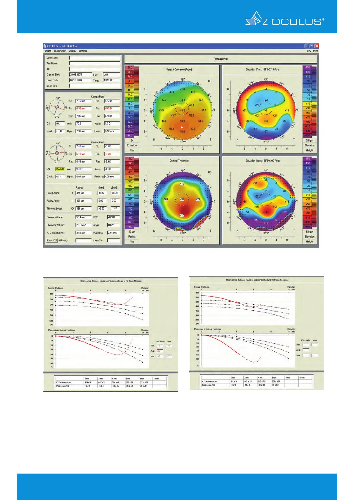
Figure 31: 4 Maps Refractive of OS showing post-LASIK ectasia
Figure 32: Pachymetry progression in OD
Figure
: 33 Pachymetry progression in OS
The pachymetric progression is abrupt in both eyes, providing a significant indication of ectasia
(Figure 32, Figure 33).
Probably mild ectasia could have been diagnosed prior to surgery if corneal topography and
tomography would have performed and well interpreted. This case would have been considered as a
bad candidate for RK.