34
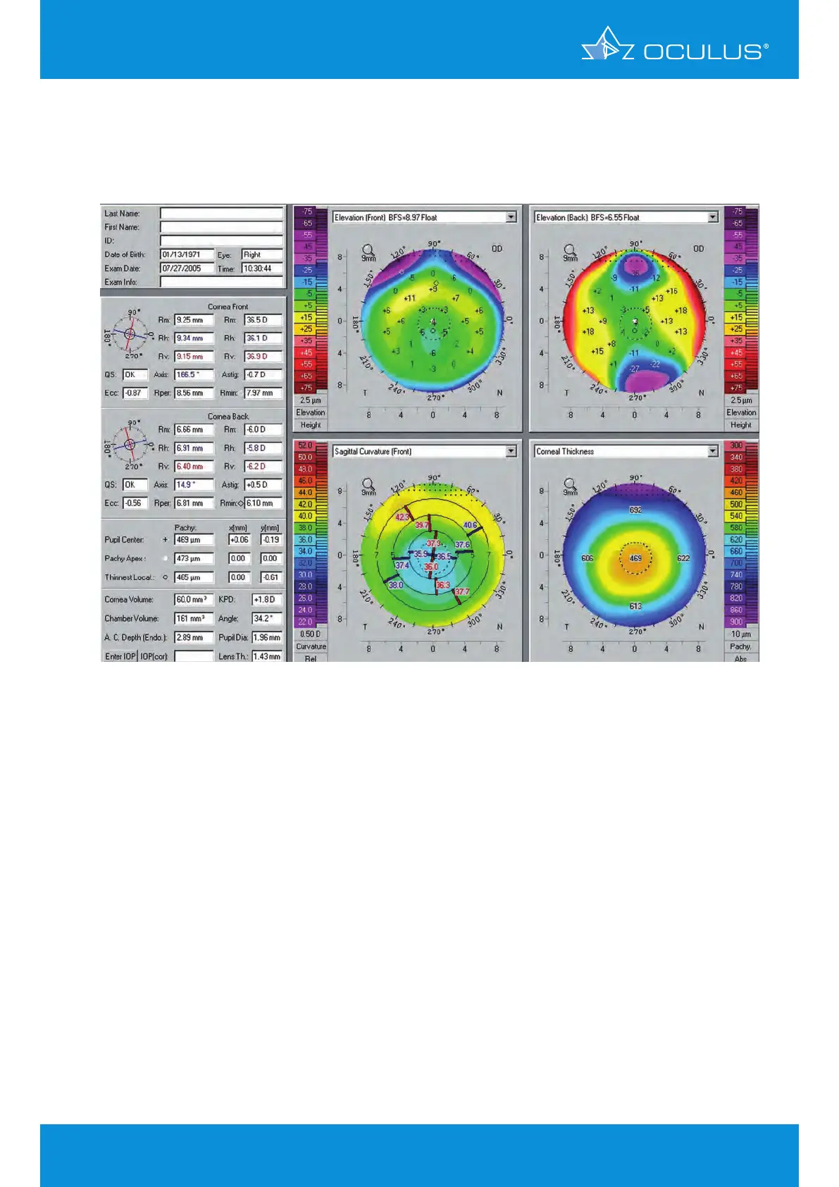
Evaluation with the Pentacam® revealed no posterior elevation abnormality and no evidence of
postoperative ectasia (Figure 35).
The patient underwent routine LASIK enhancement without incident.
Note:
This case demonstrates one of the limitations with the current version of the Bausch & Lomb
Orbscan®. This device routinely fails to correctly identify the posterior corneal surface in
postoperative patients, leading to underestimates of residual bed thickness and frequent incorrect
diagnosis of post-LASIK ectasia.
Here the Orbscan® incorrectly reads the corneal thickness 37 μm thinner than the Pentacam®,
incorrectly suggesting ectasia (Figure 36). The Pentacam® shows a normal postoperative
appearance (Figure 37).
Figure 35: 4 Maps Selectable revealing there to be no post-LASIK ectasia
8 Corneal ectasia