EasyManuals Logo

OCULUS Pentacam Interpretation Guide

OCULUS Pentacam
216 pages
To Next Page IconTo Next Page
To Previous Page IconTo Previous Page
Page #89 background imageLoading...
Page #89 background image
87
The enhanced reference surface more closely resembles the more normal periphery (Figure 108) and
allows for easier identification of ectatic regions.
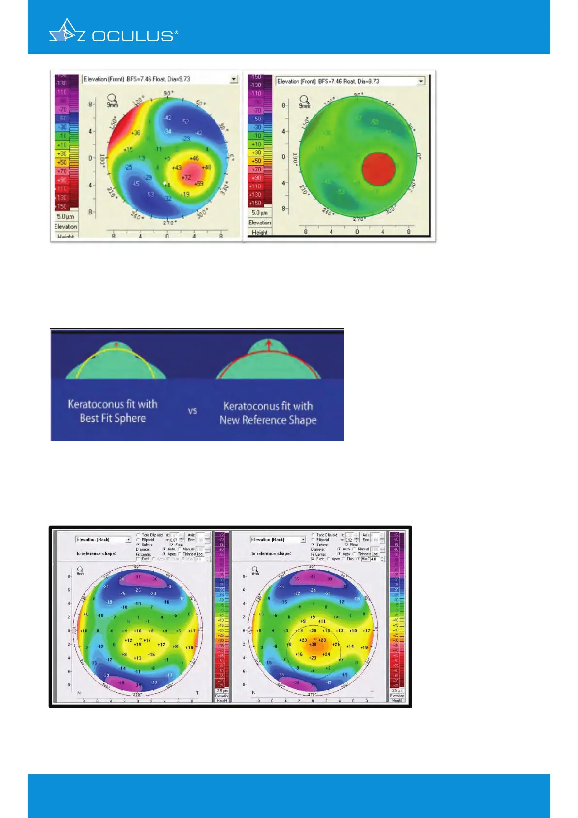
In Figure 109, the standard BFS is shown on the left, while the enhanced reference surface on the
right accentuates the ectatic region, yielding an island of greater magnitude.
The enhanced reference surface is one component of the Belin/Ambrósio Enhanced Ectasia Display,
a comprehensive tool for preoperative refractive surgery screening.
Figure 107: Elevation map of a keratoconic cornea using an enhanced reference shape
with an exclusion zone to improve detectability
Figure 109: Standard BFS on the left, enhanced reference surface on the right
Figure 108: Enhanced reference surface
12 Belin/Ambrósio Enhanced Ectasia Display

Table of Contents

Other manuals for OCULUS Pentacam

Questions and Answers:

Question and Answer IconNeed help?

Do you have a question about the OCULUS Pentacam and is the answer not in the manual?

OCULUS Pentacam Specifications

General IconGeneral
Measurement TimeLess than 2 seconds
Corneal CoverageUp to 12 mm
SoftwarePentacam Software
Data TransferUSB
Corneal TopographyYes
DisplayExternal monitor required
Measurement PrincipleScheimpflug imaging
CameraRotating Scheimpflug camera
Light SourceBlue LED
PachymetryYes
Anterior Chamber AnalysisYes
Imaging PrincipleScheimpflug imaging
FixationInternal
Power Supply100-240 V, 50/60 Hz
ResolutionHigh resolution
Lens Density MeasurementYes
DimensionsVaries depending on model and configuration
WeightVaries by model, check manufacturer specifications
Number of Images25 or 50

Related product manuals