Chapter 2 - Introduction
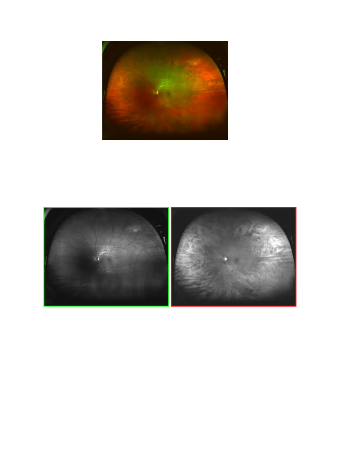
Sample image from 200Tx device.
The composite image supports analysis of the retinal image by allowing individual review of the green and
red channel information.
l
The Green Channel image contains information from the sensory retina through the pigment
epithelium layers of the retina.
l
The Red Channel image contains information from the deeper structures of the retina, from the
pigment epithelium through the choroid.
Compare the green and red channels derived from the composite image shown above.
Sample image from 200Tx device.
Green Channel view Red Channel view
The diagram below summarizes the retinal layers and structures reached by the lasers. These laser
wavelengths penetrate the retinal structures to different depths, each wavelength providing information for
interpretation and diagnosis.
4 of 100