Chapter 2 - Introduction
Image types
There are different types of image for each type of procedure:
l
optomap® – captures a retinal image. The standard optomap® procedure is a wellness exam. This
procedure captures a standard optomap® retinal image.
l
optomap® plus – captures a medical retinal image. The optomap® plus procedure is a medical
retinal exam. Following this procedure will allow the use of the enhanced features in the Review
application.
l
optomap® fa – some systems are capable of capturing fluorescein images. The optomap® fa
medical procedure allows the capture of fluorescein optomap® fa images. Following this
procedure will allow the use of the enhanced features in the Review application. These images are
captured at a higher resolution than standard optomap® images. The image capture rate for the
early-phase images can be set in the Capture application settings. You must ensure that the
necessary resources are available to administer the fluorescein.
Note
l
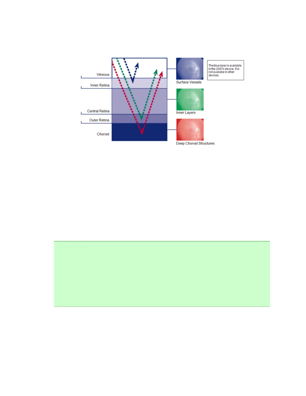
Some systems are capable of capturing autofluorescence images, How to capture
Autofluorescence images on page 27.optomap® af images are captured using a green laser
light for illumination. This contrasts with the traditional use of blue illumination or white light for
autofluorescence imaging. Green light autofluorescence differs, and allows a different
visualization of macular pathologies since macular pigment does not interfere. Also the Optic
Nerve Head is clearly visible when green laser light is used for autofluorescence imaging.
l
Some image types offer additional options, for example, eye steering, ResMax™, images
optimized for the periphery or central pole. These options vary depending on the device being
used and the image type selected. All available options are displayed in the Procedure Selection
dialog box.
5 of 100