80
I
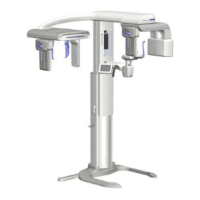
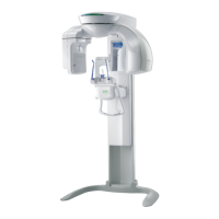
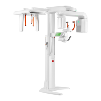
PaX-i3D Smart
7. Acquiring CEPH images
To acquire images, 5. Getting Started should be completed first. If 5. Getting Started
is not completed, return to that section and finish the step first.
❚
CEPH Imaging Software
Examination Image Description
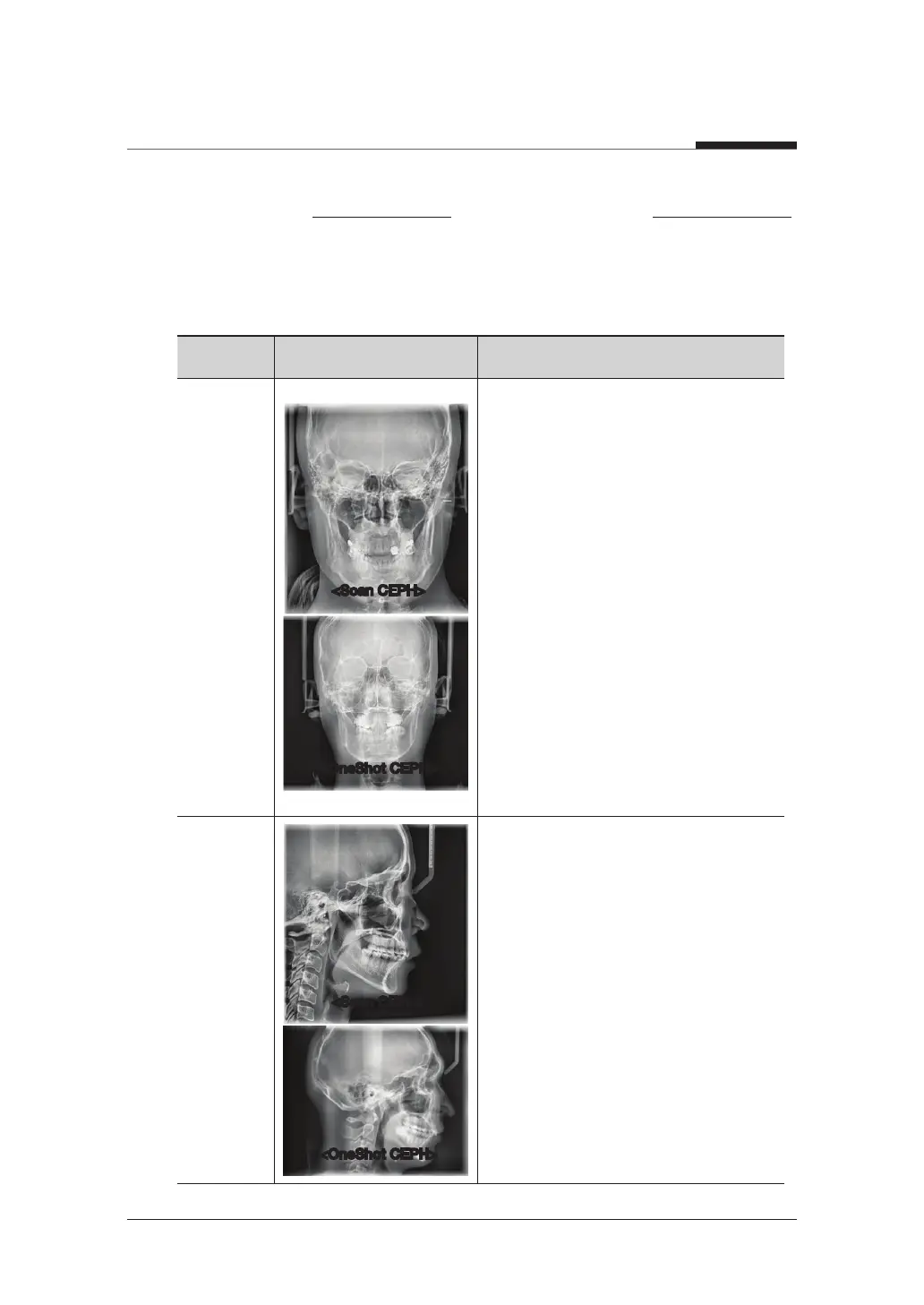
PA
● The radiation is directed from the
posterior of the skull to the anterior.
● Use to examine cranial diseases,
trauma and congenital malformations.
● Used to assess the growth of lateral
side of the face. It is also useful to
examine the ramus mandibulae, the
posterior region of the third largest
molar in the lower jaw, and the side
wall of the maxillary sinus, and the
frontal sinus, antrum ethmoidale,
olfactory pits and optic disc pits.
● Measure the angles formed by the
connecting lines between the cranial
measurement points to further assess
the growth of the facial region. It is
widely used in Orthodontics and Oral
and Maxillofacial Surgery.
Lateral
● Study craniofacial disease, trauma
and congenital malformation and
examine the soft tissue in the
otorhinolaryngological area, sinus and
hard palate.
● Measure the angles formed by the
connecting lines between the cranial
measurement points to further assess
the growth of the facial region. It's
widely used in Orthodontics and Oral
and Maxillofacial Surgery.
<Scan CEPH>
<OneShot CEPH>
<Scan CEPH>
<OneShot CEPH>